. 2012 Nov 2;287(45):38379-89.
doi: 10.1074/jbc.M112.374926. Epub 2012 Sep 19.

The obestatin/GPR39 system is up-regulated by muscle injury and functions as an autocrine regenerative system

Affiliations

The obestatin/GPR39 system is up-regulated by muscle injury and functions as an autocrine regenerative system

Uxía Gurriarán-Rodríguez et al. J Biol Chem. .

Abstract

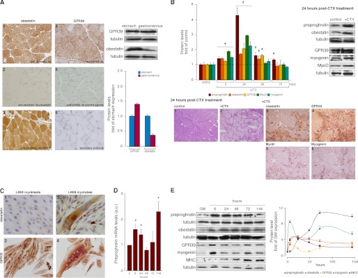
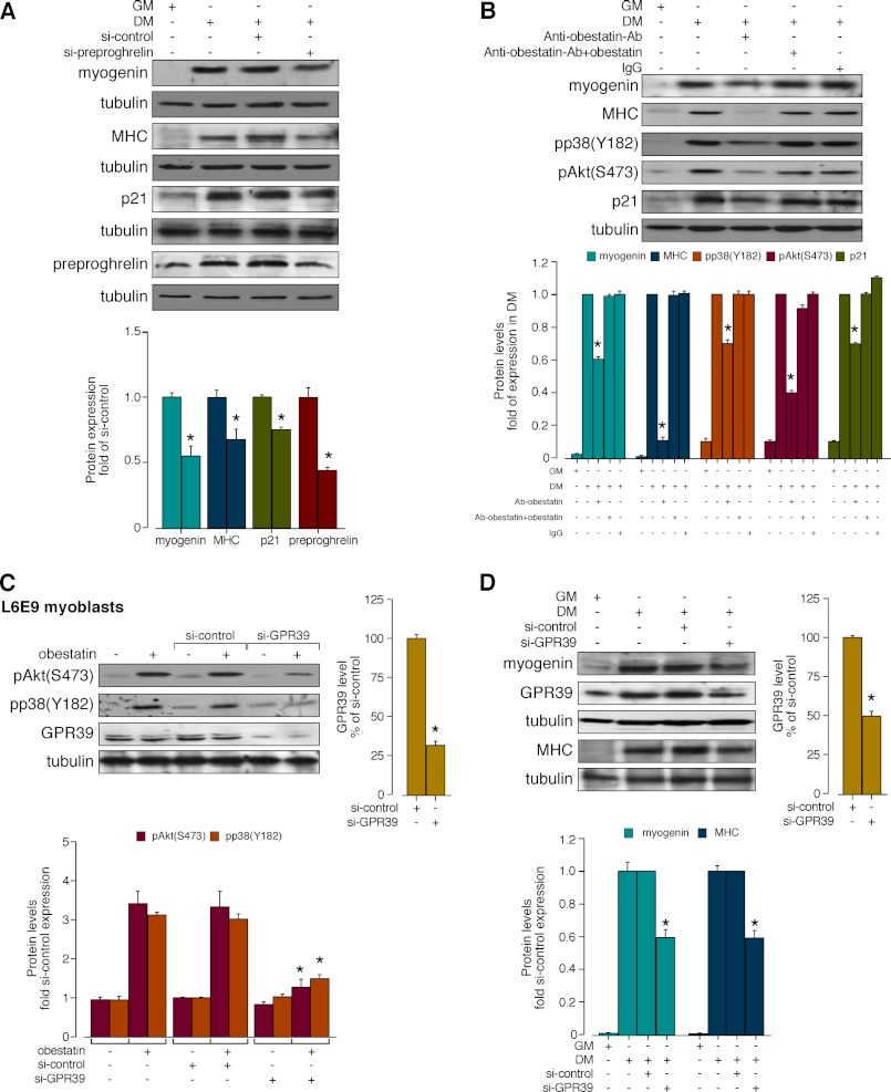
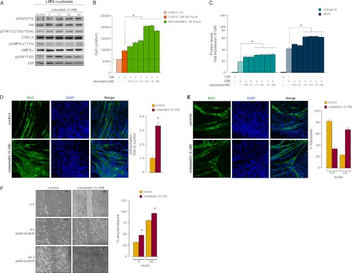
The maintenance and repair of skeletal muscle are attributable to an elaborate interaction between extrinsic and intrinsic regulatory signals that regulate the myogenic process. In the present work, we showed that obestatin, a 23-amino acid peptide encoded by the ghrelin gene, and the GPR39 receptor are expressed in rat skeletal muscle and are up-regulated upon experimental injury. To define their roles in muscle regeneration, L6E9 cells were used to perform in vitro assays. For the in vivo assays, skeletal muscle tissue was obtained from male rats and maintained under continuous subcutaneous infusion of obestatin. In differentiating L6E9 cells, preproghrelin expression and correspondingly obestatin increased during myogenesis being sustained throughout terminal differentiation. Autocrine action was demonstrated by neutralization of the endogenous obestatin secreted by differentiating L6E9 cells using a specific anti-obestatin antibody. Knockdown experiments by preproghrelin siRNA confirmed the contribution of obestatin to the myogenic program. Furthermore, GPR39 siRNA reduced obestatin action and myogenic differentiation. Exogenous obestatin stimulation was also shown to regulate myoblast migration and proliferation. Furthermore, the addition of obestatin to the differentiation medium increased myogenic differentiation of L6E9 cells. The relevance of the actions of obestatin was confirmed in vivo by the up-regulation of Pax-7, MyoD, Myf5, Myf6, myogenin, and myosin heavy chain (MHC) in obestatin-infused rats when compared with saline-infused rats. These data elucidate a novel mechanism whereby the obestatin/GPR39 system is coordinately regulated as part of the myogenic program and operates as an autocrine signal regulating skeletal myogenesis.


Figures

FIGURE 1.
FIGURE 1.
A, immunohistochemical detection of obestatin (A.1) and GPR39 (A.4) in gastrocnemius muscle (objective magnification 20×). Preadsorption controls were performed, applying the primary antibody plus obestatin (A.2), ghrelin (A.3), or GPR39 control peptide (A.5) (10 nmol/ml per control peptide) to positive samples. Immunohistochemistry was also performed in the absence of primary antibody (A.6). Right panel, immunoblot analysis of obestatin and GPR39 of extracts from stomach and gastrocnemius muscle of control rats (n = 5). Results were expressed as a -fold of protein levels in the stomach. B, preproghrelin, obestatin, and GPR39 expression in the CTX model of gastrocnemius muscle injury. Left panel, relative protein expression of preproghrelin, obestatin, GPR39, MyoD, and myogenin in CTX- or PBS-injected gastrocnemius muscle was assessed at the indicated time points by immunoblot (n = 5 per group and time point). Protein expression was expressed as a -fold over PBS control muscle. Right panel, representative immunoblot images of gastrocnemius muscle sections 24 h after PBS or CTX injection. Lower panel, representative images of gastrocnemius muscle sections 24 h after PBS or CTX injection: hematoxylin/eosin (B1, control, and B2, +CTX); immunohistochemical detection of obestatin, GPR39, MyoD, and myogenin after CTX injection (B3, B4, B5, and B6, respectively; objective magnification 20×). C, immunocytochemical detection of obestatin and GPR39 in myoblast and myotube L6E9 cells (objective magnification 40×). D, relative mRNA transcripts of preproghrelin in the course of L6E9 myogenesis. mRNA was quantified by real-time PCR and expressed as arbitrary units (n = 3). E, immunoblot analysis of preproghrelin, obestatin, GPR39, myogenin, and MHC expression in the course of L6E9 myogenesis. Protein level was expressed as a -fold over control cells in GM (n = 3). Immunoblots in A and B are representative of the mean value. Immunoblots in E are representative of three independent experiments. Data were expressed as the mean ± S.E. obtained from intensity scans of independent experiments. *, p < 0.05 versus control values.
FIGURE 2.
FIGURE 2.
A, effect of siRNA depletion of preproghrelin on myogenesis. L6E9 cells were transfected with preproghrelin siRNA prior to induction of myogenesis for 96 h. Expression of myogenin, MHC, p21, and preproghrelin was denoted as a -fold of the respective levels in control siRNA-transfected cells (n = 3). B, autocrine role of obestatin on myogenesis. Differentiating L6E9 cells were maintained in DM, DM supplemented with anti-obestatin antibody (5 μg/μl; anti-obestatin Ab), obestatin + anti-obestatin Ab (1:1, w/w), or preimmune IgG (5 μg/μl) for 6 days. Expression of pp38(Tyr-182), pAkt(Ser-473), or p21 is expressed as a -fold of respective expressions in DM control (n = 6). C, effect of GPR39 knockdown by siRNA on obestatin-activated pAkt(Ser-473) and pp38(Tyr-182) in L6E9 myoblast cells (5 nm, 5 min). GPR39 was expressed as a -fold of its level to control siRNA-transfected cells (n = 3). Activation of Akt and p38 was expressed relative to control siRNA-transfected cells. D, effect of siRNA depletion of GPR39 on myogenesis. L6E9 cells were transfected with GPR39 siRNA prior to induction of myogenesis for 96 h. Expression of myogenin and MHC was expressed as a -fold of the respective levels to control siRNA-transfected cells (n = 3). Immunoblots in A, B, C, and D are representative of three independent experiments. Data were expressed as the mean ± S.E. obtained from intensity scans of independent experiments. *, p < 0.05 versus control values.
FIGURE 3.
FIGURE 3.
A, time course of the effect of obestatin (5 nm) in L6E9 myoblast cells on pAkt(Ser-473), pERK1/2(Thr-202/Tyr-204), pAMPK(Thr-172), and pp38(Tyr-182) (n = 3). AMPK, AMP-activated protein kinase. B, dose-response effect of obestatin (0.01–100 nm) on L6E9 myoblast cell proliferation (48 h, n = 5). Co: cells initially seeded. Control: cells maintained 48 h in GM. C, dose-response effect of obestatin (0.01–100 nm) on differentiating L6E9 cells for 6 days (n = 6). Levels of myogenin and MHC were represented as a -fold of respective expression in GM. D, left panel, immunofluorescence detection of MHC and DAPI (objective magnification 20×) in L6E9 myotube cells under DM (control) or DM + obestatin (5 nm) at the 6-day point after stimulation. Right panel, the differentiation grade was evaluated based on the number of MHC-positive cells above total nuclei and expressed as a -fold of control. E, representative images used to determined the number of nuclei within individual myotubes (at least two nuclei) in L6E9 myotube cells under DM (control) or DM + obestatin (5 nm) at the 6-day point after stimulation (left panel, objective magnification 20×). Myotubes were grouped into two categories, and the percentage of myotubes in each category was determined (right panel). F, left panel, L6E9 myoblast cells at 100% confluence were wounded with a sterile pipette tip to remove cells. Photographs were taken (objective magnification 10×) at 0, 8, and 24 h after injury. Right panel, wound closure was evaluated using the equation described under “Experimental Procedures.” Immunoblots in A and C are representative of three and six independent experiments, respectively. For these panels, data were expressed as the mean ± S.E. obtained from intensity scans of independent experiments. For panels D and E, data were expressed as the mean ± S.E. as described under “Experimental Procedures.” *, p < 0.05 versus control values.
FIGURE 4.
FIGURE 4.
Effect of 72-h continuous subcutaneous infusion of obestatin (300 nmol/kg of body weight/24 h; n = 10) on myogenic markers (MyoD, Myf5, Pax-7, myogenin, Myf6, and MHC) and myofiber cross-section area (A) and VEGF, VEGFR2, and PEDF expression (B). Protein levels were expressed as a -fold of control obtained from a 72-h minipump subcutaneously implanted in saline group (n = 10 per group). Immunoblots in A and B are representative of mean values from each rat group. In A, hematoxylin/eosin-stained gastrocnemius (right, upper panel) and soleus (right, lower panel) sections are representative of the mean value of total cross-section area. Data were expressed as the mean ± S.E. *, p < 0.05 versus control values.

Similar articles

Cited by

References

    1. Chargé S. B., Rudnicki M. A. (2004) Cellular and molecular regulation of muscle regeneration. Physiol. Rev. 84, 209–238 - PubMed
    1. Tedesco F. S., Dellavalle A., Diaz-Manera J., Messina G., Cossu G. (2010) Repairing skeletal muscle: regenerative potential of skeletal muscle stem cells. J. Clin. Invest. 120, 11–19 - PMC - PubMed
    1. Kuang S., Gillespie M. A., Rudnicki M. A. (2008) Niche regulation of muscle satellite cell self-renewal and differentiation. Cell Stem Cell 2, 22–31 - PubMed
    1. Bentzinger C. F., Wang Y. X., Rudnicki M. A. (2012) Building muscle: molecular regulation of myogenesis Cold Spring Harb. Perspect. Biol. 4, pii: a008342 - PMC - PubMed
    1. DiMario J., Buffinger N., Yamada S., Strohman R. C. (1989) Fibroblast growth factor in the extracellular matrix of dystrophic (mdx) mouse muscle. Science 244, 688–690 - PubMed

Publication types

MeSH terms