. 2013 Feb;133(2):344-53.
doi: 10.1038/jid.2012.329. Epub 2012 Sep 20.

Tbx18 targets dermal condensates for labeling, isolation, and gene ablation during embryonic hair follicle formation

Affiliations

Tbx18 targets dermal condensates for labeling, isolation, and gene ablation during embryonic hair follicle formation

Laura Grisanti et al. J Invest Dermatol. 2013 Feb.

Abstract

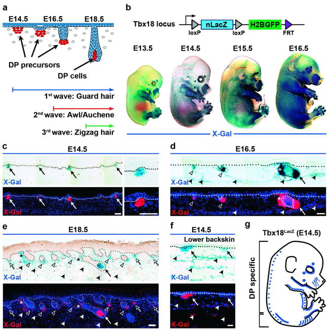
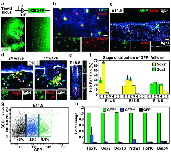
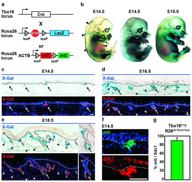
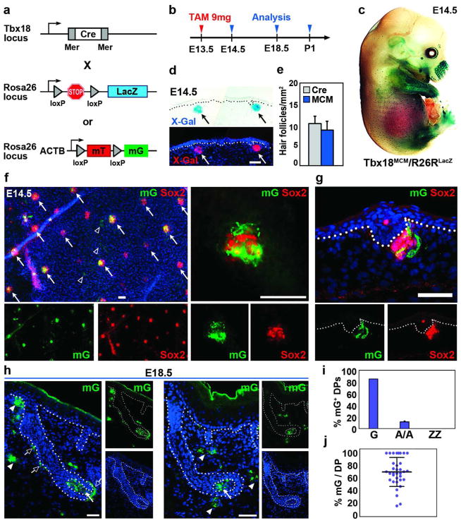
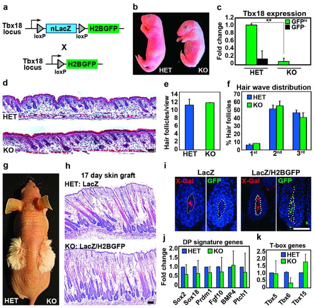
How cell fate decisions of stem and progenitor cells are regulated by their microenvironment or niche is a central question in stem cell and regenerative biology. Although functional analysis of hair follicle epithelial stem cells by gene targeting is well established, the molecular and genetic characterization of the dermal counterpart during embryonic morphogenesis has been lacking because of the absence of cell type-specific drivers. Here, we report that T-box transcription factor Tbx18 specifically marks dermal papilla (DP) precursor cells during embryonic hair follicle morphogenesis. With Tbx18(LacZ), Tbx18(H2BGFP), and Tbx18(Cre) knock-in mouse models, we demonstrate LacZ and H2BGFP (nuclear green fluorescent protein) expression and Cre activity in dermal condensates of nascent first-wave hair follicles at E14.5. As Tbx18 expression becomes more widespread throughout the dermis at later developmental stages, we use tamoxifen-inducible Cre-expressing mice, Tbx18(MerCreMer), to exclusively target DP precursor cells and their progeny. Finally, we ablate Tbx18 in full knockout mice, but find no perturbations in hair follicle formation, suggesting that Tbx18 is dispensable for normal DP function. In summary, our study establishes Tbx18 as a genetic driver to target for the first time embryonic DP precursors for labeling, isolation, and gene ablation that will greatly enhance investigations into their molecular functions during hair follicle morphogenesis.


Conflict of interest statement

CONFLICT OF INTEREST

The authors state no conflict of interest.

Figures

Figure 1
Figure 1. Tbx18LacZ expression in embryonic skin
(a) Schematic of embryonic hair follicle development. Dermal condensates are visible at E14.5. (b) Top: Schematic of Tbx18LacZ reporter. Bottom: Whole-mount X-Gal staining in embryos. (c–f) Top: LacZ expression in skin sections at different ages. Bottom: Pseudo-colored X-Gal (red) overlaid with Dapi to highlight nuclei. The dotted line marks basement membrane. (c) LacZ in dermal condensates at E14.5 (arrows). (d) LacZ expression in DP cells of downgrowing guard hair follicles at E16.5 (arrow) and in second-wave DPs (open arrowheads). (e) At E18.5, all DPs express LacZ in downgrowing follicles (arrow, open arrowheads) and third wave dermal condensates (open arrows). (d,e) Note weak LacZ expression in dermis (filled arrowheads) and arrector pili muscle (asterisks). (f) Weak LacZ expression in dermal cells in lower backskin at E14.5 (arrowheads). (g) Schematic summarizing Tbx18 expression in embryonic skin at E14.5. Blue dots illustrate Tbx18 expression in DP precursor cells, the blue line widespread expression in the dermis (lower bracket). Bars = 50 μm.
Figure 2
Figure 2. Labeling and isolation of dermal condensates with Tbx18H2BGFP
(a) Schematic of Tbx18H2BGFP reporter. Below E14.5 embryo (left) showing GFP expression in hair follicles. (b–e) Sox2 co-localization with GFP in DP precursors and more mature DPs. Whole-mount skin (b) and sagittal section (c, arrows) at E14.5. (d) GFP expression in first- and second-wave DP follicles (arrows) and dermis (arrowheads) at E16.5. (e) GFP and Sox2 co-expression in mature DP. Itgb4 marks basement membrane. Dapi highlights all nuclei (blue). Bar = 50μm. (f) Quantification of Sox2 cells in GFP-positive DPs in all developmental stages (n=4). (g) FACS isolation of DP precursors at E14.5. Three cell populations were isolated: GFPhi, dermal condensates (green); GFPlow, dermal cells from lower backskin (blue); GFP, negative cells (black). (h) Real-time PCR analysis of isolated cells. Only GFPhi cells were enriched in dermal condensate genes. Data shown are mean ± SD (n=2).
Figure 3
Figure 3. Tbx18Cre activity in DP precursor cells during hair follicle formation
(a) Schematic of Tbx18Cre, R26RLacZ and R26ACTB-mT/mG reporter lines. (b) Whole-mount X-Gal staining of Tbx18Cre/R26RLacZ embryos at E14.5 showed Cre activity in a hair follicle pattern. Few labeled follicles were visible at E14.0 (arrowheads). (c) Cre activity in DP precursor cells at E14.5 (arrows). (d,e) At E16.5 and E18.5, Cre activity is present in all DP cells (arrows, open arrowheads) and dermal condensates (open arrows), and more widespread in the dermis and arrector pili muscle (filled arrowheads, asterisks). (f) Immunofluorescence for Sdc1 (Syndecan1) confirmed identity of GFP positive dermal condensates with the R26ACTB-mT/mG reporter. (g) Quantification of GFP and Sdc1 double-labeled dermal condensates (n=2). Bars = 50μm. Data shown are mean ± SD.
Figure 4
Figure 4. Inducible Cre activity in Tbx18MerCreMer mice targets guard hair DP cells and their progeny
(a) Schematic of tamoxifen (TAM) inducible Tbx18MerCreMer (MCM) mice crossed with R26RLacZ or R26ACTB-mT/mG reporters. (b) Timeline of Cre induction and reporter analysis. (c) Whole-mount X-gal staining of Tbx18MCM/R26RLacZ embryo showed reporter activation in hair follicle pattern at E14.5. (d) Inducible Cre activity in dermal condensates. (e) Quantification of labeled follicles in Tbx18Cre and Tbx18MCM embryos. (f) Co-expression of Sox2 and GFP in DPs of Tbx18MCM/R26ACTB-mT/mG whole-mount skin at E14.5 (arrows). Few dermal cells were labeled (arrowheads); asterisks mark autofluorescence; right: high magnification. (g) Co-localization in sagittal section. (h) GFP expression in DP (arrows) and dermal sheath cells (open arrows) of guard hair follicles at E18.5. Few dermal cells were GFP positive (arrowheads). (i) Quantification of follicles with GFP-positive DP cells (n=3). (j) Quantification of GFP-positive DP cells per DP in embryo sections. In ~25% of guard hairs 100% DP cells were labeled (n=3). Bar = 50μm. Data shown are mean ± SD.
Figure 5
Figure 5. Tbx18 in DP precursor cells is not required for hair follicle formation
(a) Schematic of generating Tbx18 knockouts. (b) Newborn Tbx18 knockout (KO) and heterozygous (HET) pups. (c) Verification of Tbx18 ablation by Real-time PCR in FACS isolated DP precursor cells at E14.5 (n=3). (d) Hematoxylin and eosin staining of newborn backskins HET and KO. (e) Unchanged total hair follicle numbers (n=3). (f) All hair follicle subtypes are formed in KO (n=3). (g) Normal hair shaft formation in skin grafts of newborn HET (Tbx18LacZ) and KO (Tbx18LacZ/H2BGFP). (h) Hematoxylin and eosin staining showed normal follicle morphologies in KO skin grafts. (i) LacZ and GFP expression in DPs confirming donor origin of grafted skins. (j,k) Real-time PCRs of FACS-isolated DPs at E14.5 for DP signature genes (j) and T-box family members (k) (n=3). Bar = 50μm. Data shown are mean ± SD. **, p<0.01.

Similar articles

Cited by

References

    1. Andl T, Reddy ST, Gaddapara T, Millar SE. WNT signals are required for the initiation of hair follicle development. Dev Cell. 2002;2:643–53. - PubMed
    1. Botchkarev VA, Botchkareva NV, Roth W, Nakamura M, Chen LH, Herzog W, et al. Noggin is a mesenchymally derived stimulator of hair-follicle induction. Nat Cell Biol. 1999;1:158–64. - PubMed
    1. Bussen M, Petry M, Schuster-Gossler K, Leitges M, Gossler A, Kispert A. The T-box transcription factor Tbx18 maintains the separation of anterior and posterior somite compartments. Genes Dev. 2004;18:1209–21. - PMC - PubMed
    1. Cai CL, Martin JC, Sun Y, Cui L, Wang L, Ouyang K, et al. A myocardial lineage derives from Tbx18 epicardial cells. Nature. 2008;454:104–8. - PMC - PubMed
    1. Chen D, Jarrell A, Guo C, Lang R, Atit R. Dermal β-catenin activity in response to epidermal Wnt ligands is required for fibroblast proliferation and hair follicle initiation. Development. 2012;139:1522–33. - PMC - PubMed

Publication types

MeSH terms

LinkOut - more resources