. 2012 Nov;139(21):3986-96.
doi: 10.1242/dev.082909. Epub 2012 Sep 19.

Pancortins interact with amyloid precursor protein and modulate cortical cell migration

Affiliations

Pancortins interact with amyloid precursor protein and modulate cortical cell migration

Heather C Rice et al. Development. 2012 Nov.

Abstract

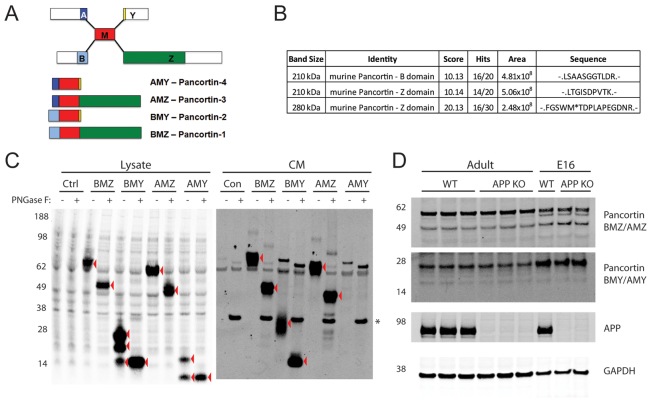
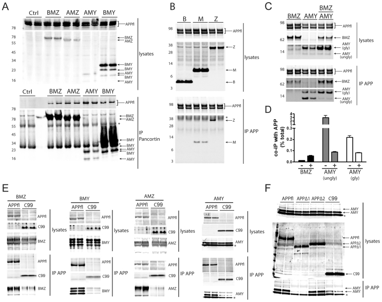
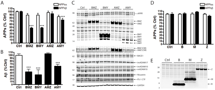
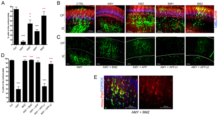
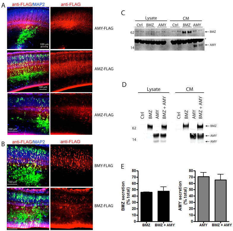
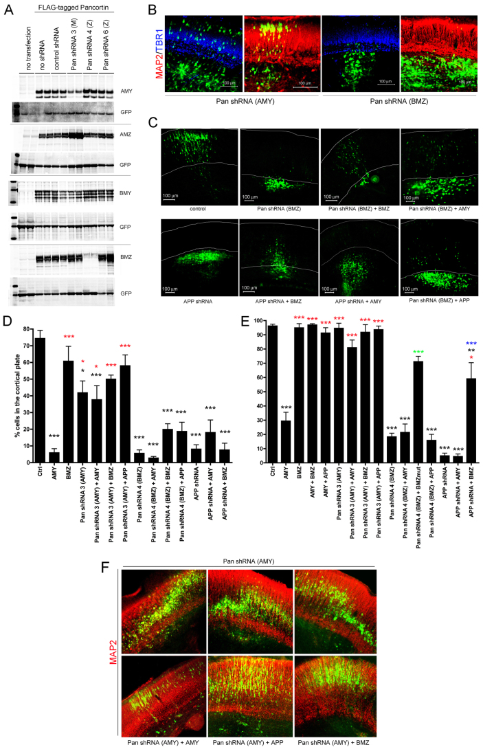
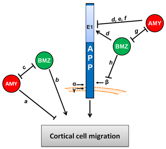
Neuronal precursor cell migration in the developing mammalian brain is a complex process requiring the coordinated interaction of numerous proteins. We have recently shown that amyloid precursor protein (APP) plays a role in migration into the cortical plate through its interaction with two cytosolic signaling proteins, disabled 1 (DAB1) and disrupted in schizophrenia 1 (DISC1). In order to identify extracellular factors that may signal through APP to regulate migration, we performed an unbiased mass spectrometry-based screen for factors that bind to the extracellular domain of APP in the rodent brain. Through this screen, we identified an interaction between APP and pancortins, proteins expressed throughout the developing and mature cerebral cortex. Via co-immunoprecipitation, we show that APP interacts with all four of the mammalian pancortin isoforms (AMY, AMZ, BMY, BMZ). We demonstrate that the BMZ and BMY isoforms of pancortin can specifically reduce β-secretase- but not α-secretase-mediated cleavage of endogenous APP in cell culture, suggesting a biochemical consequence of the association between pancortins and APP. Using in utero electroporation to overexpress and knock down specific pancortin isoforms, we reveal a novel role for pancortins in migration into the cortical plate. Interestingly, we observe opposing roles for alternate pancortin isoforms, with AMY overexpression and BMZ knock down both preventing proper migration of neuronal precursor cells. Finally, we show that BMZ can partially rescue a loss of APP expression and that APP can rescue effects of AMY overexpression, suggesting that pancortins act in conjunction with APP to regulate entry into the cortical plate. Taken together, these results suggest a biochemical and functional interaction between APP and pancortins, and reveal a previously unidentified role for pancortins in mammalian cortical development.


Figures

Fig. 1.
Fig. 1.
Identification of pancortin as an APP-binding partner. (A) Schematic of pancortin isoforms and their domains. (B) Pancortin peptides identified by mass spectrometry in an unbiased screen for extracellular factors within murine cortical slices that interact with the APP ectodomain. (C) Western blot (WB) for pancortin (anti-FLAG) of the lysate and conditioned media (CM) from HEK293 cells transiently transfected with each of the pancortins or control vector both with and without PNGase F treatment. Red arrowheads highlight specific bands. (D) Western blot for pancortin isoforms (‘BMZ/AMZ’ and ‘BMY/AMY’), APP and GAPDH in E16 and adult brain homogenates from wild-type or APP knockout mice.
Fig. 2.
Fig. 2.
APP and pancortins biochemically interact. Western blots for APP (anti-APP, C9) and FLAG-tagged pancortin (anti-FLAG, M2) are shown for both the input (lysates) and the immunoprecipitated products. Immunoprecipitations of lysates for FLAG (M2) are shown in A and for APP (C9) in B-F. Asterisks represent non-specific or cross-reactive IgG bands. (A) Western blots of lysates of HEK293-APP695 cells transiently transfected with FLAG-tagged pancortin isoforms or vector alone (ctrl). (B) Western blots of lysates and IPs of HEK293-APP695 cells transiently transfected with FLAG-tagged pancortin domains B, M and Z. (C) Western blots of lysates and IPs of HEK293-APP695 cells transiently transfected with BMZ and AMY isoforms alone or together. (D) Quantification of the percent of BMZ and AMY co-immunoprecipitated with APP relative to the total amount of each isoform in the lysate for BMZ and both the unglycosylated (lower band, ‘ungly’) and glycosylated (upper band, ‘gly’) forms of AMY when BMZ and AMY were expressed separately (−) or together (+). (E) Western blots of lysates and IPs of HEK293 cells transiently transfected with each of the pancortins and either full-length APP (APPfl) or a C-terminal fragment of APP (C99). (F) Western blots of lysates and IPs of HEK293 cells transiently transfected with the AMY isoform of pancortin and either full-length APP, APPΔ1 (lacking residues 36-289), APPΔ2 (lacking residues 288-493) or C99.
Fig. 3.
Fig. 3.
Specific pancortin isoforms reduce β-secretase- but not α-secretase-mediated cleavage of APP. HEK293 cells were transiently transfected with pancortin isoforms and individual domains or empty vector (ctrl). Twenty-four hours post-transfection, media were replaced, and after 24 additional hours media were collected and cells lysed. (A,D) Quantification of endogenous APPsα and APPsβ in the conditioned media (CM) via a multiplex ELISA, which allows for the simultaneous detection of each. (B) Quantification of Aβ40 in the CM by a separate ELISA. (C,E) Western blots of lysates (and conditioned media where noted by ‘CM’) showing expression of endogenous full-length APP, transfected pancortins and each of the secretases. Asterisk in C indicates a non-specific band. In A,B,D, each bar represents data from two to four independent experiments performed in triplicate for A and B, and a single representative experiment performed in triplicate for D. Error bars represent s.e.m. ***P<0.001.
Fig. 4.
Fig. 4.
Expression levels of different pancortin isoforms have opposing effects in migration of neural precursor cells into the cortical plate. E15.5 rat embryos were co-electroporated with GFP and constructs encoding pancortin cDNAs. (A,D) Quantification of the percent of GFP-positive cells that migrated into the cortical plate after 3 (A) and 6 (D) days post electroporation. Each bar represents the average of data acquired from at least three independent embryos electroporated with constructs listed. Error bars represent s.e.m. *P<0.05, ***P<0.005. Red and black asterisks indicate significance relative to AMY and control, respectively. (B) Representative images from control brains or those expressing the listed pancortin isoforms 3 days post-electroporation. MAP2 immunostaining is shown in red and TBR1 in blue. CP, cortical plate; IZ, intermediate zone. (C) Representative images showing the 6 days post-electroporation time point following electroporation of AMY with and without BMZ or APP co-electroporation. White lines indicate boundaries of the cortical plate. (E) Two panels of a representative image showing GFP, pancortin immunostaining (red) and TOPRO-3 staining (blue), or pancortin immunostaining alone of sections electroporated with AMY and BMZ.
Fig. 5.
Fig. 5.
Differential secretion of the pancortin isoforms from neuronal cells. (A,B) E15.5 rat embryos were co-electroporated in utero with GFP and constructs listed, and harvested 3 days later. Brains were sectioned and immunostained for FLAG and MAP2. Shown are representative images of GFP (green), anti-FLAG (red) and anti-MAP2 (blue). (C,D) Lysates and CM from ex vivo cultures of cortical neurons electroporated in utero (C) or transfected in vitro (D). BMZ or AMY were immunoprecipitated with M2 (anti-Flag)-agarose and western blots performed to detect FLAG-tagged pancortin (anti-FLAG) (C,D). (E) Quantification of the percent of BMZ and AMY detected in the CM relative to total (CM + lysate) following in vitro transfection of BMZ and AMY both together and separately.
Fig. 6.
Fig. 6.
Pancortins are required for proper migration in the developing cerebral cortex. (A) shRNA constructs targeting pancortin isoforms were generated and tested for their ability to knock down each of the FLAG-tagged pancortin isoforms. HEK293 cells were transfected with construct combinations as labeled. GFP was co-transfected in all cases to monitor transfection efficiency. Forty-eight hours following transfection, cells were lysed and western blots performed to examine GFP and pancortin levels, using M2 (α-FLAG). (B-F) E15.5 rat embryos were electroporated in utero with GFP and constructs listed, and harvested 3 or 6 days later. Brains were dissected, fixed, sectioned coronally and immunostained for MAP2 and TBR1. (B) Representative images of a 3-day harvest following introduction of shRNA 3 (which knocks down AMY) and shRNA 4 (which knocks down BMZ). MAP2 immunostaining is shown in red and TBR1 in blue. (C) Representative images of brains electroporated with constructs listed and harvested 6 days later. White lines delineate the boundaries of the cortical plate as determined by MAP2 and TBR1 immunostaining. (D,E) Quantification of the percentage of GFP-positive cells present in the cortical plate 3 (D) and 6 (E) days post electroporation. Each bar presents data from the average of at least three independent embryos electroporated with constructs listed. Error bars represent s.e.m. *P<0.05, **P<0.01, ***P<0.005. Black asterisks represent significance relative to GFP vector control; red asterisks represent significance relative to AMY; green asterisks represent significance relative to pancortin shRNA 4 (BMZ); blue asterisks represent significance relative to APP shRNA. Quantifications from Fig. 3A,D for GFP, AMY, BMZ, AMY+BMZ and AMY+APP are re-shown here for direct comparison with knock down data. (F) Representative images of brains electroporated with shRNA targeting AMY with and without co-electroporation of AMY, APP or BMZ and harvested at 6 days post electroporation. MAP2 immunostaining is shown in red.
Fig. 7.
Fig. 7.
Summary of the effects observed of pancortins and APP on migration in the developing cerebral cortex. In utero electroporation of pancortin constructs in the developing rat brain revealed novel and opposing roles of the AMY and BMZ isoforms in cortical cell migration. (a) Cells overexpressing AMY fail to enter the cortical plate, suggesting that AMY inhibits cortical plate entry. (b) Knock down of BMZ results in failure to enter the cortical plate, suggesting that endogenous BMZ promotes cortical plate entry. (c) BMZ overexpression rescues the defect observed with AMY overexpression. Taken together with previous studies of neuronal differentiation in Xenopus (Moreno and Bronner-Fraser, 2005), the data support a model where AMY and BMZ mutually inhibit the activity of the other. (d) In support of an APP-dependent mechanism, peptides to the B and Z domain of pancortin were identified in an unbiased mass spectrometry screen for APP interacting proteins and both BMZ and AMY co-immunoprecipitate with APP, an interaction disrupted by deletion of the E1 region of the APP ectodomain. (e,f) Overexpression of APP rescues AMY overexpression (e), but blockade of the interaction through deletion of the AMY binding site within APP (the E1 domain) (f) prevents rescue of the AMY defect by APP, supporting a model whereby AMY inhibits the function of APP in cortical cell migration through a physical interaction. (g) Expression of BMZ can decrease the co-immunoprecipitation efficiency of AMY with APP, which suggest that BMZ may rescue the AMY defect by preventing AMY from binding to APP. (h) BMZ inhibits β-secretase cleavage of APP; however, whether this activity is mechanistically involved in regulating migration has yet to be determined.

References

    1. Araki W., Kitaguchi N., Tokushima Y., Ishii K., Aratake H., Shimohama S., Nakamura S., Kimura J. (1991). Trophic effect of β-amyloid precursor protein on cerebral cortical neurons in culture. Biochem. Biophys. Res. Commun. 181, 265-271 - PubMed
    1. Bai Y., Markham K., Chen F., Weerasekera R., Watts J., Horne P., Wakutani Y., Bagshaw R., Mathews P. M., Fraser P. E., et al. (2008). The in vivo brain interactome of the amyloid precursor protein. Mol. Cell. Proteomics 7, 15-34 - PubMed
    1. Barembaum M., Moreno T. A., LaBonne C., Sechrist J., Bronner-Fraser M. (2000). Noelin-1 is a secreted glycoprotein involved in generation of the neural crest. Nat. Cell Biol. 2, 219-225 - PubMed
    1. Bulfone A., Smiga S. M., Shimamura K., Peterson A., Puelles L., Rubenstein J. L. (1995). T-brain-1: a homolog of Brachyury whose expression defines molecularly distinct domains within the cerebral cortex. Neuron 15, 63-78 - PubMed
    1. Cheng A., Arumugam T. V., Liu D., Khatri R. G., Mustafa K., Kwak S., Ling H. P., Gonzales C., Xin O., Jo D. G., et al. (2007). Pancortin-2 interacts with WAVE1 and Bcl-xL in a mitochondria-associated protein complex that mediates ischemic neuronal death. J. Neurosci. 27, 1519-1528 - PMC - PubMed

Publication types

MeSH terms