. 2012 Dec;86(23):12826-37.
doi: 10.1128/JVI.00917-12. Epub 2012 Sep 19.

Ataxia telangiectasia mutated kinase controls chronic gammaherpesvirus infection

Affiliations

Ataxia telangiectasia mutated kinase controls chronic gammaherpesvirus infection

Joseph M Kulinski et al. J Virol. 2012 Dec.

Abstract

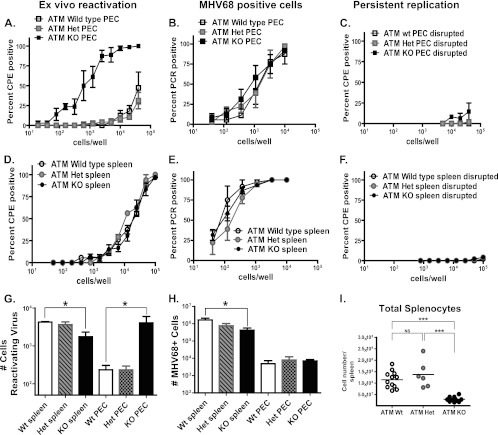
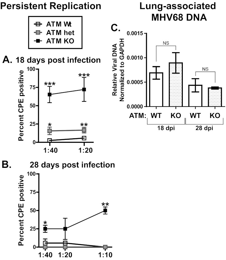
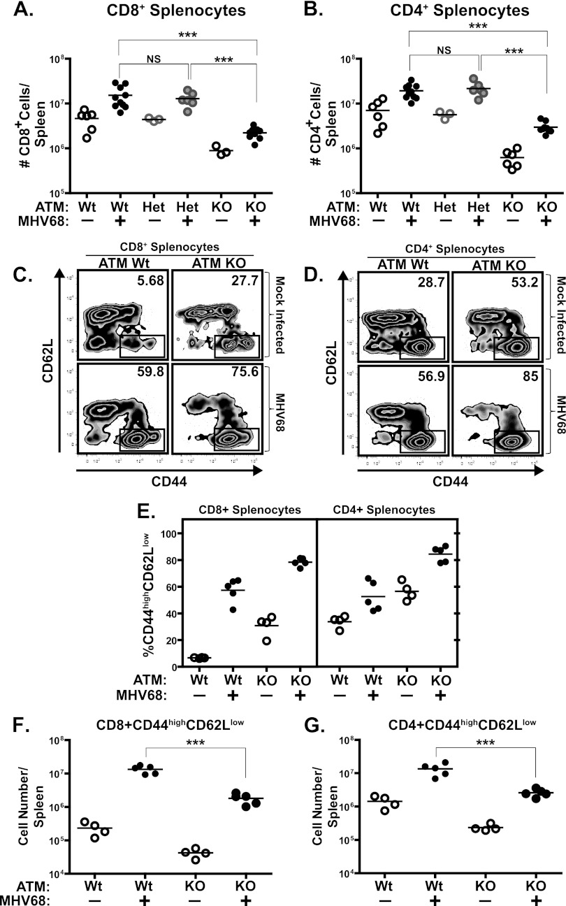
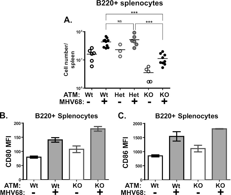
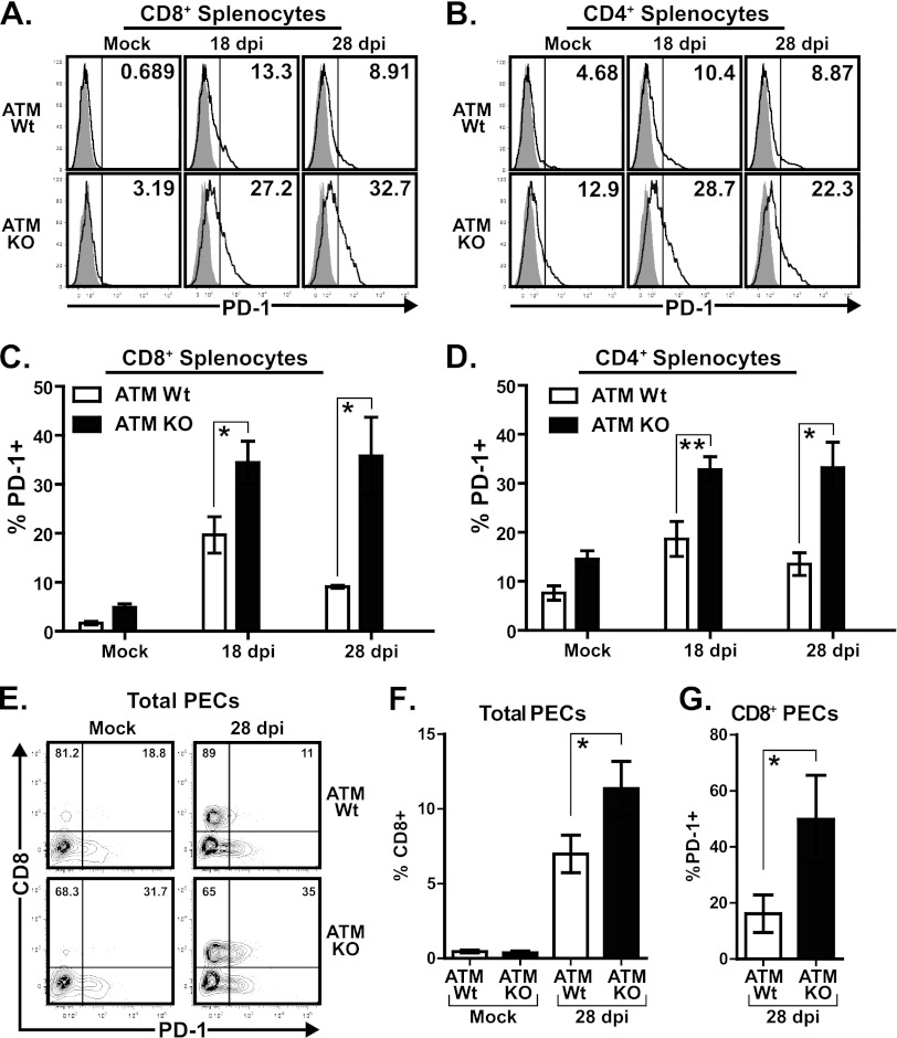
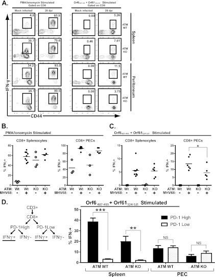
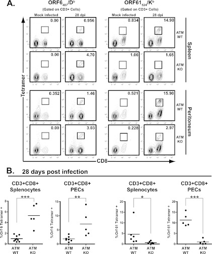
Gammaherpesviruses, such as Epstein-Barr virus (EBV), are ubiquitous cancer-associated pathogens that interact with DNA damage response, a tumor suppressor network. Chronic gammaherpesvirus infection and pathogenesis in a DNA damage response-insufficient host are poorly understood. Ataxia-telangiectasia (A-T) is associated with insufficiency of ataxia-telangiectasia mutated (ATM), a critical DNA damage response kinase. A-T patients display a pattern of anti-EBV antibodies suggestive of poorly controlled EBV replication; however, parameters of chronic EBV infection and pathogenesis in the A-T population remain unclear. Here we demonstrate that chronic gammaherpesvirus infection is poorly controlled in an animal model of A-T. Intriguingly, in spite of a global increase in T cell activation and numbers in wild-type (wt) and ATM-deficient mice in response to mouse gammaherpesvirus 68 (MHV68) infection, the generation of an MHV68-specific immune response was altered in the absence of ATM. Our finding that ATM expression is necessary for an optimal adaptive immune response against gammaherpesvirus unveils an important connection between DNA damage response and immune control of chronic gammaherpesvirus infection, a connection that is likely to impact viral pathogenesis in an ATM-insufficient host.


Figures

Fig 1
Fig 1
MHV68 latency is inadequately controlled in ATM-deficient mice. ATM-deficient (KO), heterozygous (Het), or wild-type mice were intranasally inoculated with 104 PFU of MHV68. Splenocytes and peritoneal exudate cells (PEC) were harvested at 18 days postinfection and pooled from 3 to 5 mice in each experimental group. (A to F) Frequencies of infected cells (B and E), ex vivo reactivation (A and D), and persistent replication (C and F) were determined using limiting-dilution assays. (G and H) Absolute numbers of reactivating cells (G) and MHV68-positive cells (H) per anatomic site were calculated using the frequencies determined in panels A, B, D, and E and absolute cell counts. (I) Numbers of splenocytes in MHV68-infected mice with the indicated genotypes at 18 days postinfection. Each symbol represents an individual spleen. For all panels, data were pooled from 3 or 4 independent experiments. *, P < 0.05; ***, P < 0.001; NS, P > 0.05.
Fig 2
Fig 2
Persistent MHV68 replication in ATM-deficient lungs. Lungs were harvested at 18 or 28 days postinfection (dpi) with 104 PFU MHV68. (A and B) Serial dilutions of lung homogenates were plated on indicator MEF monolayers and the presence of lytic virus scored in each replicate after 14 days of culture. Data were pooled from 10 wild-type mice, 8 ATM-heterozygous, and 6 ATM KO mice at 18 days postinfection and from 3 mice per experimental group at 28 days postinfection. *, P < 0.05; **, P < 0.01; ***, P < 0.001 (for each indicated dilution compared to the wild-type group). (C) DNA was isolated from lung homogenates and subjected to qPCR analysis for quantification of MHV68-specific DNA. Data represent the mean and standard error from analysis of 4 mice per experimental group at 18 days postinfection and 3 mice per group at 28 days postinfection. NS, P > 0.05.
Fig 3
Fig 3
T cell numbers and activation status in ATM-deficient mice. ATM-deficient, heterozygous, or wild-type mice were intranasally inoculated with 104 PFU of MHV68 or carrier solution. Splenocytes were harvested at 18 days postinfection and stained with either anti-CD4 or anti-CD8 antibody (A and B) or in a tristain cocktail with the addition of anti-CD44 and anti-CD62L (C to G). Panels C and D are profiles of CD8- or CD4-gated splenocytes from individual spleens and are representative of at least 2 independent experiments (values in upper right corners indicate the percentage of CD44high CD62Llow splenocytes). Panels A, B, and E to G represent pooled data from 2 or 3 independent experiments, where floating bars represent the mean and each symbol represents an individual spleen. *, P < 0.05; **, P < 0.01; ***, P < 0.001; NS, P > 0.05.
Fig 4
Fig 4
B cell numbers and activation status in ATM-deficient mice. ATM-deficient, heterozygous, or wild-type mice were intranasally inoculated with 104 PFU of MHV68 or sterile carrier solution (mock). (A) Absolute numbers of B220+ splenocytes at 18 days postinfection. Each symbol represents an individual spleen; data were pooled from 2 or 3 independent experiments. (B and C) Mean fluorescent intensity of CD80 and CD86 staining of B220+ splenocytes at 18 days postinfection. Data were pooled from 2 or 3 independent experiments. *, P < 0.05; **, P < 0.01; ***, P < 0.001; NS, P > 0.05.
Fig 5
Fig 5
T cells of MHV68-infected ATM-deficient mice increase PD-1 expression. ATM-deficient or wild-type mice were intranasally inoculated with 104 PFU of MHV68 or carrier solution (mock). Splenocytes or peritoneal exudate cells (PEC) were harvested at 18 or 28 days postinfection (dpi), stained with anti-PD1 in combination with either anti-CD8 or CD4 antibodies, and analyzed by flow cytometry. (A and B) Representative histograms show PD-1 gating of each experimental group (shaded, isotype control; solid line, anti-PD-1). Values in the upper right corners correspond to the percentage of CD8+ or CD4+ splenocytes positive for anti-PD-1 staining as determined by the gate shown (vertical line). (C and D) Percentage of CD4- or CD8-positive cells expressing cell surface PD-1 using the gate shown in panels A and B. Data are pooled from 3 to 5 mice per group from 3 independent experiments and are shown as the mean and standard error. (E) Representative flow cytometry plots showing anti-CD8 and anti-PD-1 staining in peritoneal exudate cells (PEC), where values in the upper left and upper right quadrants correspond to the percentage of CD8+ PEC that are either negative or positive for PD-1, respectively. (F and G) Pooled data from at least 4 independent experiments (4 to 6 mice per group) showing the percentage of total PEC that are positive for CD8 (F) or the percentage of CD8+ PEC that are positive for PD-1 (G) based on the gates shown in panel E. *, P < 0.05; **, P < 0.01.
Fig 6
Fig 6
Fewer peritoneal CD8+ T cells from infected ATM-deficient mice produce IFN-γ upon MHV68 peptide restimulation. ATM-deficient or wild-type mice were intranasally inoculated with 104 PFU of MHV68 or carrier solution (mock infected). Splenocytes or peritoneal exudate cells (PEC) were harvested at 28 days postinfection (dpi) and restimulated ex vivo with either PMA-ionomycin or ORF6487-495 plus ORF61524-531 peptide for 5 h before surface staining with anti-CD8, anti-CD44, and anti-PD-1 and intracellular staining with anti-IFN-γ for analysis by flow cytometry. (A) Representative flow cytometry plots showing CD44 and IFN-γ staining of CD8+-gated cells, where values in the upper right corners represent the percentage of CD8+ cells within the gate shown. (B and C) Combined data from at least 4 independent experiments showing the percentage of CD8+ cells positive for intracellular IFN-γ staining based on the gating strategy shown in panel A. Floating bars represent the mean, and each symbol represents an individual mouse. (D) Pooled data from at least 4 independent experiments (4 to 6 mice per experimental group) showing the mean and standard error of the percentage of CD8+ PD-1-high or CD8+ PD-1-low populations that produce IFN-γ upon ex vivo restimulation with MHV68 peptides (as determined by the gating strategy shown in the schematic). *, P < 0.05; **, P < 0.01; ***, P < 0.001; NS, P > 0.05.
Fig 7
Fig 7
ATM-deficient mice exhibit a skewed proportion of MHV68-specific CD8+ T cells. ATM-deficient or wild-type mice were intranasally inoculated with 104 PFU of MHV68 or carrier solution (mock infected). Splenocytes or peritoneal exudate cells (PEC) were harvested at 28 days postinfection (dpi) and stained with antibodies against CD3 and CD8 in combination with either ORF6487/Db or ORF61524/Kb MHC class I tetramers for analysis by flow cytometry. (A) Representative flow cytometry plots showing anti-CD8 and MHC class I tetramer staining of CD3+-gated cells, where values in the upper right corner of each plot represent the percentage of CD3+CD8+ cells within the gate shown. (B) Pooled data from at least 4 independent experiments showing the percentage of CD3+ CD8+ cells positive for MHC class I tetramer staining based on the gating strategy shown in panel A. Floating bars represent the mean, and each symbol represents an individual mouse. *, P < 0.05; **, P < 0.01; ***, P < 0.001.

Comment in

Similar articles

Cited by

References

    1. Barlow C, et al. 1996. Atm-deficient mice: a paradigm of ataxia telangiectasia. Cell 86:159–171 - PubMed
    1. Barton E, Mandal P, Speck SH. 2011. Pathogenesis and host control of gammaherpesviruses: lessons from the mouse. Annu. Rev. Immunol. 29:351–397 - PubMed
    1. Barton ES, Lutzke ML, Rochford R, Virgin HW. 2005. Alpha/beta interferons regulate murine gammaherpesvirus latent gene expression and reactivation from latency. J. Virol. 79:14149–14160 - PMC - PubMed
    1. Berkel AI, et al. 1979. Epstein-Barr virus-related antibody patterns in ataxia-telangiectasia. Clin. Exp. Immunol. 35:196–201 - PMC - PubMed
    1. Brooks JW, et al. 1999. Requirement for CD40 ligand, CD4(+) T cells, and B cells in an infectious mononucleosis-like syndrome. J. Virol. 73:9650–9654 - PMC - PubMed

Publication types

MeSH terms

Substances