. 2012 Dec;86(23):12871-8.
doi: 10.1128/JVI.01981-12. Epub 2012 Sep 19.

Overexpression of the ubiquitin-specific protease 7 resulting from transfection or mutations in the ICP0 binding site accelerates rather than depresses herpes simplex virus 1 gene expression

Affiliations

Overexpression of the ubiquitin-specific protease 7 resulting from transfection or mutations in the ICP0 binding site accelerates rather than depresses herpes simplex virus 1 gene expression

Maria Kalamvoki et al. J Virol. 2012 Dec.

Abstract

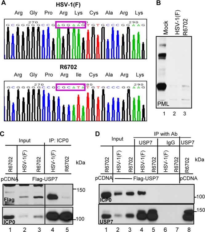
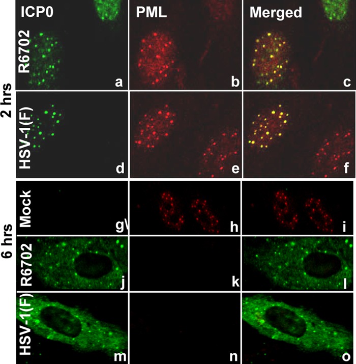
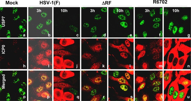
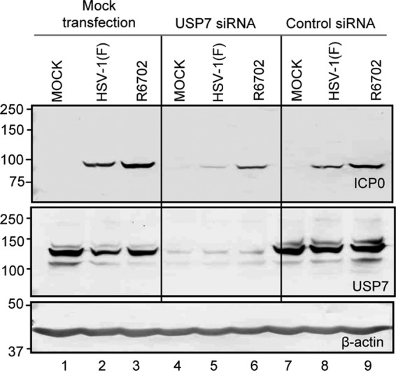
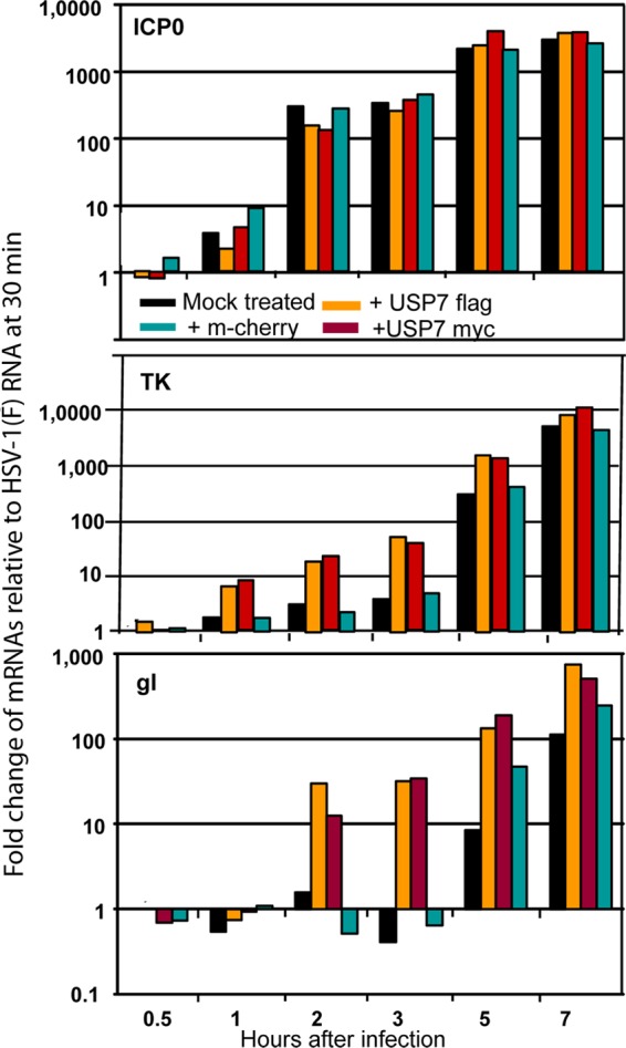
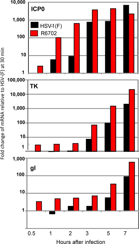
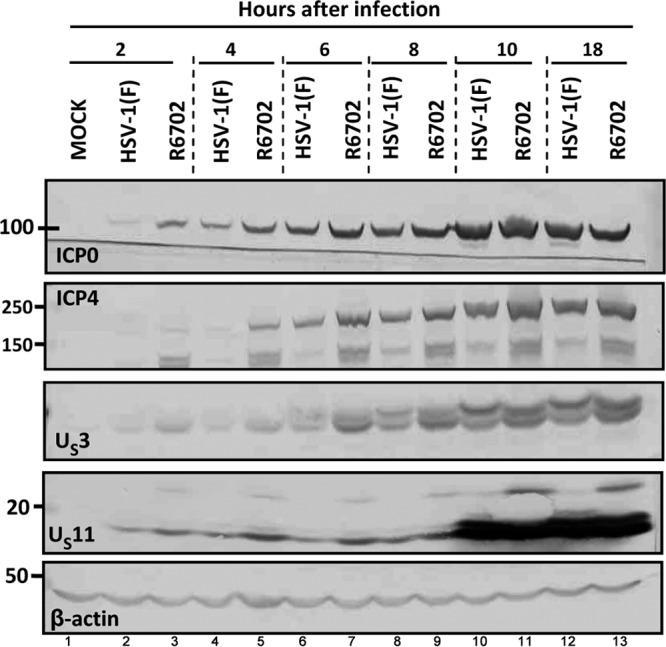
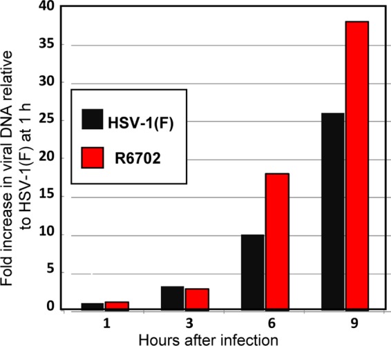
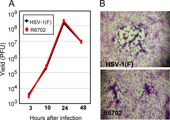
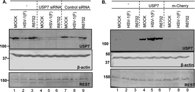
Earlier studies reported that ICP0, a key regulatory protein encoded by herpes simplex virus 1 (HSV-1), binds ubiquitin-specific protease 7 (USP7). The fundamental conclusion of these studies is that depletion of USP7 destabilized ICP0, that ICP0 mediated the degradation of USP7, and that amino acid substitutions in ICP0 that abolished binding to USP7 significantly impaired the ability of HSV-1 to replicate. We show here that, indeed, depletion of USP7 leads to reduction of ICP0 and that USP7 is degraded in an ICP0-dependent manner. However, overexpression of USP7 or substitution in ICP0 of a single amino acid to abolish binding to USP7 accelerated the accumulation of viral mRNAs and proteins at early times after infection and had no deleterious effect on virus yields. A clue as to why USP7 is degraded emerged from the observation that, notwithstanding the accelerated expression of viral genes, the plaques formed by the mutant virus were very small, implying a defect in virus transmission from cell to cell.


Figures

Fig 1
Fig 1
Construction and characterization of a USP7 binding-defective recombinant virus, R6702. (A) Sequence chromatographs of HSV-1(F) and R6702. The mutation from a lysine residue in HSV-1(F) into an isoleucine residue in R6702 is highlighted by purple rectangles. (B) Viral protein expression and PML degradation of R6702. pHEL cells were infected with HSV-1(F) or R6702 at 5 PFU/cell. At 6 h postinfection, the cells were lysed, and total cell lysates were probed with various antibodies, as indicated. (C and D) Interaction between ICP0 and USP7 in HSV-1(F) and R6702. HEp-2 cells transfected with Flag-tagged USP7 or pcDNA3.1 were infected with HSV-1(F) or R6702. At 18 h postinfection, cells were harvested, washed, and lysed by brief sonication. Total cell lysates were cleared and reacted with either anti-ICP0 antibody (Ab) (C) or anti-USP7 antibody (D) for immunoprecipitation (IP). Rabbit IgG (IgG) was used as a negative control. The immunoprecipitates were probed for USP7, Flag, or ICP0 as indicated.
Fig 2
Fig 2
R6702 ICP0 localization is similar to that of wt ICP0. pHEL cells seeded in 4-well slides were infected with 10 PFU of either HSV-1(F) (d to f and m to o) or R6702 (a to c and j to l) per cell. The cells were fixed at 2 h (a to f) or at 6 h (j to o) after exposure to the viruses, permeabilized, and reacted simultaneously with the rabbit polyclonal antibody to ICP0 and the mouse monoclonal antibody to PML. Fluorescein isothiocyanate (FITC)-conjugated anti-rabbit and Texas Red-conjugated anti-mouse antibodies were used as secondary antibodies. The images were taken with an LSM 410 Zeiss confocal microscope.
Fig 3
Fig 3
R6702 ICP0 does not degrade USP7. HEp-2 cells seeded in 4-well slides were either mock infected (a, h, and o) or exposed to 10 PFU per cell of either HSV-1(F) (b, c, i, j, p, and q), RF (d, e, k, l, r, and s), or R6702 (f, g, m, n, t, and u). The cells were fixed at 3 h (b, i, p, d, k, r, f, m, and t) or at 10 h (c, j, q, g, n, u, a, h, o, e, l, and s) after exposure to the viruses, permeabilized, and reacted simultaneously with the rabbit polyclonal antibody to ICP0 and the mouse monoclonal antibody to USP7. Alexa-Fluor 594-conjugated goat anti-rabbit or Alexa-Fluor 488-conjugated goat anti-mouse was used as a secondary antibody. The images were taken with an LSM 410 Zeiss confocal microscope.
Fig 4
Fig 4
Knockdown of USP7 and its effect on ICP0 accumulation. HEp-2 cells were mock transfected or transfected with either USP7 siRNA or nontarget control siRNA for 96 h before being mock infected or infected with HSV-1(F) or R6702 for 10 h. Cells were harvested, lysed, and immunoblotted with the antibodies indicated. Equal amounts of HEp-2 cells that were infected without any prior treatment (lanes 1 to 3) served as controls.
Fig 5
Fig 5
USP7 overexpression increases the rates of HSV-1(F) gene transcription. HEK-293 cells seeded in 6-well plates were transfected either with USP7 Flag- or with USP7 myc-expressing plasmids. Transfections with an mCherry-expressing plasmid or nontransfected cells served as controls. The cells were infected with 3 PFU of HSV-1(F) per cell at 48 h posttransfection. Samples were collected 30 min, 1 h, 2 h, 3 h, 5 h, and 7 h after exposure to the virus, and total RNA was extracted and converted to cDNA as described in Materials and Methods; equal amounts of the cDNA were used as templates for quantification of the viral gene transcripts ICP0, TK, and gI. 18s rRNA was used for the normalization process. The results represent the fold change of the viral gene transcripts relative to the respective HSV-1(F) mRNA in mock-treated cells at 30 min after infection.
Fig 6
Fig 6
Accelerated viral gene transcription in R6702-infected cells. HEp-2 cells seeded in 6-well plates were exposed to 5 PFU of either HSV-1(F) or R6702 per cell. Samples were harvested at 30 min, 1 h, 2 h, 3 h, 5 h, or 7 h after infection and processed for total RNA isolation, cDNA synthesis, and viral gene transcript quantification, as described in Materials and Methods. The results represent the fold change of the viral gene transcripts relative to the respective HSV-1(F) mRNA at 30 min after infection.
Fig 7
Fig 7
Early accumulation of viral gene products in R6702-infected cells. HEp-2 cells were either mock infected or exposed to 5 PFU of HSV-1(F) or R6702 per cell. Samples were harvested at 2 h, 4 h, 6 h, 8 h, 10 h, or 18 h after infection, and approximately 60 μg total proteins per sample was electrophoretically separated on 10% denaturing polyacrylamide gels, electrically transferred to nitrocellulose sheets, and immunoblotted with ICP0, ICP4, US3, or US11 antibodies. β-Actin served as a loading control.
Fig 8
Fig 8
Viral DNA synthesis in R6702-infected cells. HEp-2 cells were exposed to 5 PFU of either HSV-1(F) or R6702 per cell. Samples were collected at 1 h, 3 h, 6 h, and 9 h after infection, and total DNA was isolated and used as the template to quantify the amount of viral DNA. Primers for the viral TK were used to identify the viral DNA. Primers for the β-actin gene were used as internal controls for the quantification process. The data represent the fold increase of the viral DNA relative to the DNA of HSV-1(F) present in the cells at 1 h after infection.
Fig 9
Fig 9
Growth properties of the R6702 mutant. (A) HEp-2 cells were exposed to 0.2 PFU of either HSV-1(F) or R6702 per cell. Samples were collected at 3 h, 10 h, 24 h, or 48 h after exposure to the viruses, and titrations were done on Vero cells. (B) Viral plaques from previous titrations on Vero cells, stained with Giemsa.
Fig 10
Fig 10
Stability of REST in USP7-depleted or USP7-enriched cells. (A) HEp-2 cells were transfected with 100 pmol of either USP7 or control nontarget siRNA for 72 h. The cultures were then exposed to 10 PFU of either HSV-1(F) or R6702 per cell. Samples were collected at 9 h after infection, and approximately 40 μg of total protein was electrophoretically separated on 9% denaturing polyacrylamide gels, electrically transferred to nitrocellulose sheets, and immunoblotted with REST or USP7 antibodies. β-Actin served as a loading control. (B) 293 cells were transfected with a USP7- or mCherry-expressing plasmid. At 48 h after transfection, the cells were exposed to 10 PFU of either HSV-1(F) or R6702 per cell. Samples were analyzed as for panel A.

Similar articles

Cited by

References

    1. Boutell C, Canning M, Orr A, Everett RD. 2005. Reciprocal activities between herpes simplex virus type 1 regulatory protein ICP0, a ubiquitin E3 ligase, and ubiquitin-specific protease USP7. J. Virol. 79:12342–12354 - PMC - PubMed
    1. Boutell C, Everett RD. 2003. The herpes simplex virus type 1 (HSV-1) regulatory protein ICP0 interacts with and ubiquitinates p53. J. Biol. Chem. 278:36596–36602 - PubMed
    1. Boutell C, Everett RD. 2004. Herpes simplex virus type 1 infection induces the stabilization of p53 in a USP7- and ATM-independent manner. J. Virol. 78:8068–8077 - PMC - PubMed
    1. Canning M, Boutell C, Parkinson J, Everett RD. 2004. A RING finger ubiquitin ligase is protected from autocatalyzed ubiquitination and degradation by binding to ubiquitin-specific protease USP7. J. Biol. Chem. 279:38160–38168 - PubMed
    1. Chee AV, Lopez P, Pandolfi PP, Roizman B. 2003. Promyelocytic leukemia protein mediates interferon-based anti-herpes simplex virus 1 effects. J. Virol. 77:7101–7105 - PMC - PubMed

Publication types

MeSH terms