. 2012 Dec;86(23):12861-70.
doi: 10.1128/JVI.01606-12. Epub 2012 Sep 19.

Insights into avian influenza virus pathogenicity: the hemagglutinin precursor HA0 of subtype H16 has an alpha-helix structure in its cleavage site with inefficient HA1/HA2 cleavage

Affiliations

Insights into avian influenza virus pathogenicity: the hemagglutinin precursor HA0 of subtype H16 has an alpha-helix structure in its cleavage site with inefficient HA1/HA2 cleavage

Xishan Lu et al. J Virol. 2012 Dec.

Abstract

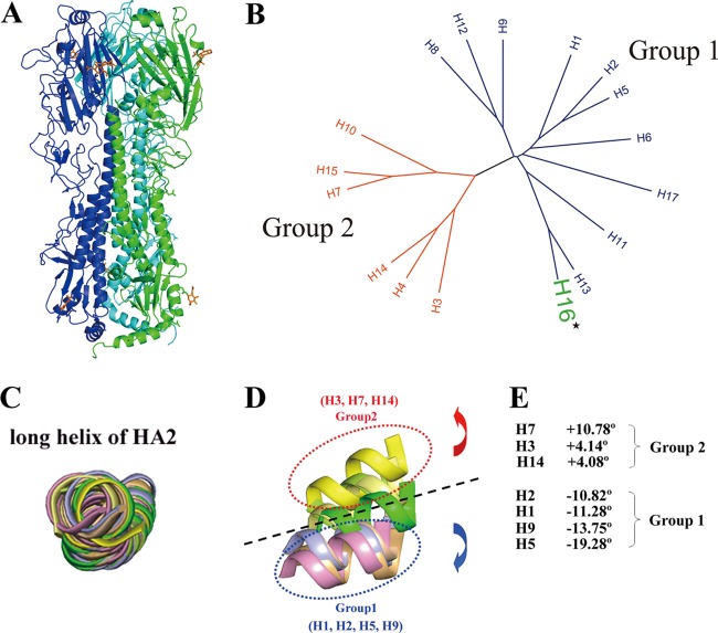
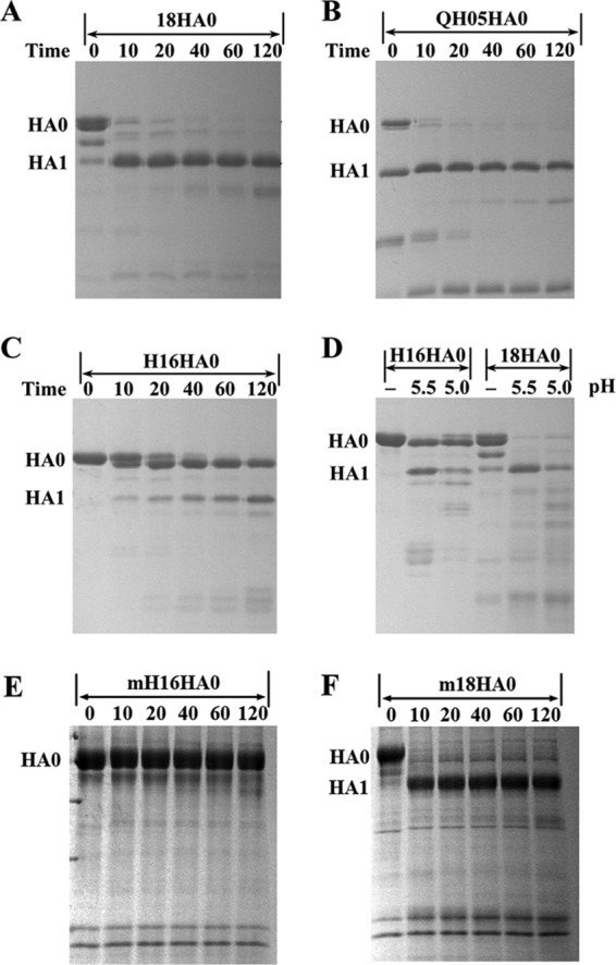
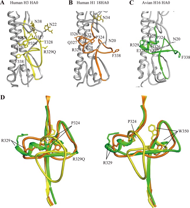
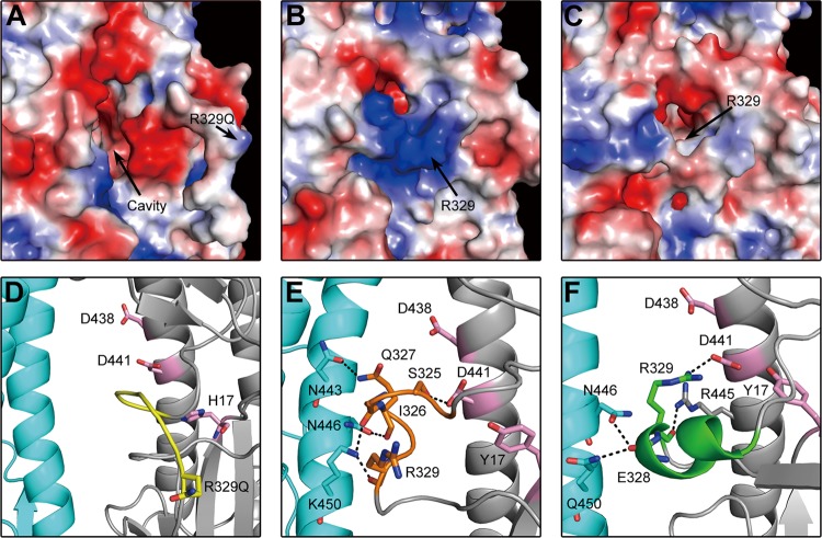
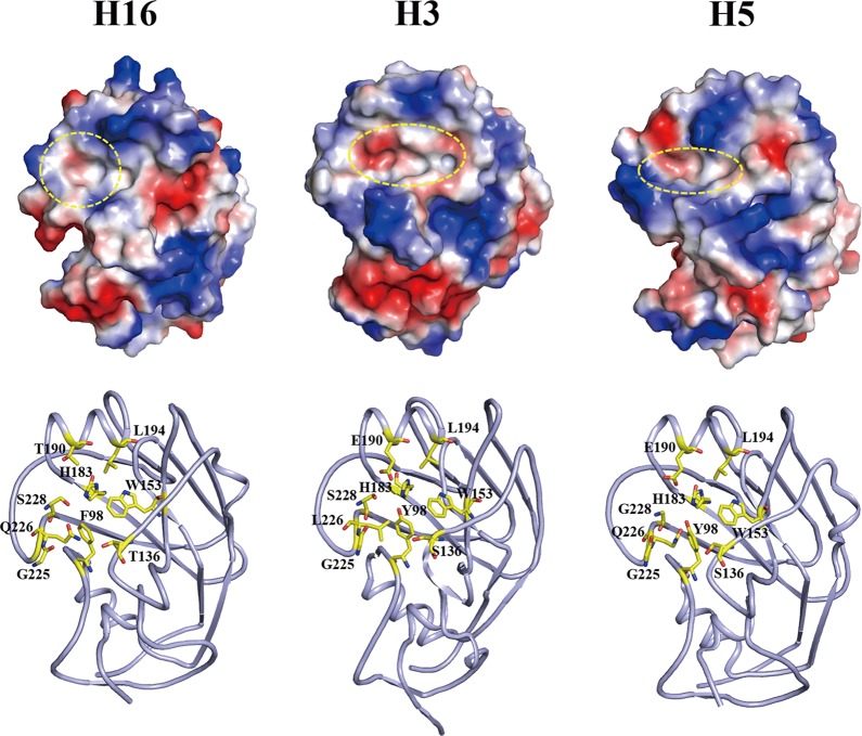
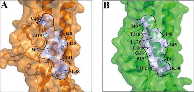
With a new serotype (H17) of hemagglutinin (HA) recently being discovered, there are now 17 serotypes (H1 to H17) of influenza A viruses in total. It is believed that HA is initially expressed as a precursor of HA0 and then cleaved into HA1 and HA2, forming a disulfide bond-linked complex, for its full function. Structural data show that a loop structure exists in the cleavage site between HA1 and HA2, and this flexible loop is crucial for the efficient cleavage of HA0. Here, the crystal structures of H16 (a low-pathogenicity avian influenza virus) in their HA0 form (H16HA0) have been solved at 1.7-Å and 2.0-Å resolutions. To our surprise, an α-helix element in the cleavage site which inserts into the negatively charged cavity with the key residue R329 hidden behind the helix was observed. In vitro trypsin cleavage experiments demonstrated inefficient cleavage of H16HA0 under both neutral and low-pH conditions. The results provide new insights into influenza A virus pathogenicity; both the relatively stable α-helix structure in the flexible cleavage loop and inaccessibility of the cleavage site likely contribute to the low pathogenicity of avian influenza A virus. Furthermore, compared to all of the HAs whose structures have been solved, H16 is a good reference for assigning the HA subtypes into two groups on the basis of the three-dimensional structure, which is consistent with the phylogenetic grouping. We conclude that in light of the current H16HA0 structure, the natural α-helix element might provide a new opportunity for influenza virus inhibitor design.


Figures

Fig 1
Fig 1
Overall structure of H16H0 and HA grouping based on 3D structures. (A) Overview of the H16HA0 trimer, represented as a ribbon diagram. For clarity, each monomer has been colored differently (A, green; B, blue; C, cyan). Carbohydrates observed in the electron density maps are colored orange. (B) Phylogenetic tree of the 17 HA subtypes. The 17 HA subtypes can be divided phylogenetically into two groups on the basis of their full-length sequences: group 1 (blue) and group 2 (red). H16, highlighted in green, belongs to group 1. (C to E) Superimpositions of the long α helix (residues 76 to 126) of HA2 reveal the displacements of the HA1 globular subdomain in seven HA subtype structures. The 190 helix (residues 188 to 195) in the receptor binding domain is used as a representative of the globular subdomain of each HA subtype. H1 is colored light blue, H2 is wheat, H3 is lemon, H5 is light orange, H7 is yellow, H9 is pink, H14 is pale yellow, and H16 is green. When we use H16 as a reference, the group 1 members rotate downward with negative-angle degrees (from −10.82° to −19.28°), while the group 2 members rotate upward with positive-angle degrees (from 4.08° to 10.78°).
Fig 2
Fig 2
Enzymatic susceptibilities of different HA0s to trypsin. Enzymatic assay with time course of from 0 to 120 min at neutral pH (8.0) for 18HA0 (A), QH05HA0 (B), and H16HA0 (C), which were produced in a baculovirus expression system. The temperature of trypsin incubation is 37°C. It is clear that QH05HA0 is a mixture of HA0 and HA1/HA2 forms when purified from the baculovirus expression system. (D) Enzymatic assay of H16HA0 and 18HA0 at low pH (5.5 and 5.0). H16HA0 is clearly resistant to the trypsin treatment. (E and F) To put this enzymatic resistance in a biological perspective, enzymatic assays of mammalian cell-expressed H16HA0 (E) and 18HA0 (F) (mH16HA0 and m18HA0, respectively) were performed. H16HA0 exhibits similar resistance to trypsin digestion, whereas 18HA0 displays similar trypsin susceptibility.
Fig 3
Fig 3
Structural comparison of the H16HA0 cleavage site with other HA0s. HA2 domains for human H3HA0 and human H1 18HA0 were aligned with H16HA0. The cleavage sites are colored yellow for human H3HA0 (A), orange for human 18HA0 (B), and green for avian H16HA0 (C). (D) Overlay of the cleavage sites of H3HA0, 18HA0, and H16HA0. The two views differ by a rotation of 90° about the 3-fold vertical axis. For H3HA0, the cleavage site forms a loop structure that projects from the glycoprotein surface, while for 18HA0, the cleavage site forms a loop structure that abuts the glycoprotein surface. Extraordinarily, in H16HA0, the cleavage site forms an α-helix structure that has not been observed before.
Fig 4
Fig 4
Sequence alignment of the cleavage sites from different H16 virus strains and a similar α-helix conformation in the cleavage site of the HA16-V327G structure. (A) Sequence alignment of the cleavage sites from 17 previously reported H16 virus strains. The most polymorphic amino acids occur at position 327, such as G, V, N, S, and D. (B) Conformation of the cleavage site in the HA16-V327G structure. Although residue V327 is replaced by glycine (G), the cleavage site still forms an α-helix structure element.
Fig 5
Fig 5
Different positions of key R329 residues in the cleavage site. (A to C) Electrostatic diagrams of cleavage sites show that the key R329 residue is far from the negatively charged cavity in H3HA0 (A), R329 covers the negatively charged cavity in 18HA0 (B), and R329 is buried in the negatively charged cavity in H16HA0 (C). (D to F) Detailed interaction between the cleavage site and the negatively charged cavity and adjacent subunit in different HA0s. The cleavage site is colored yellow in H3HA0 (D), orange in 18HA0 (E), and green in H16HA0 (F). The main residues in the negatively charged cavity are colored pink. The hydrogen bonds are shown as black dashed lines.
Fig 6
Fig 6
Structural comparison of the HA receptor binding site. (Top) Electrostatic-potential maps of the head domains of the H16 structure, human H3 structure (PDB accession number 1HA0), and avian H5 structure (PDB accession number 2FK0) were generated by the PyMOL program. The shape of the receptor binding site is marked by a yellow circle. (Bottom) Cartoon diagrams of the receptor binding site are shown with key residues that determine the specificity. Clearly, the H16HA0 receptor binding site possesses a round cavity, whereas the H3 and H5 HAs have an oval-shaped cavity. The H5 HA has a narrower cavity than the H3 HA, which is unfavorable for the human receptor binding. A broader cavity and avian-specific residue Q226 indicate that H16HA0 likely binds to both the human and avian receptors.
Fig 7
Fig 7
The conserved hydrophobic groove in H16HA0 reveals the structural basis of binding with the broadly neutralizing antibody FI6. Surface representations of the F subdomains of 09H1 HA (A) and H16HA0 (B) with selected side chains that contribute to the conserved hydrophobic groove are shown. The approximate boundaries of the hydrophobic grooves are indicated by the black lines. Although the residues contributing to the hydrophobic groove are moderately different between 09H1 and H16, similar hydrophobic grooves guarantee the binding potential by the FI6 antibody.

Similar articles

Cited by

References

    1. Anonymous 1994. The CCP4 suite: programs for protein crystallography. Acta Crystallogr. D Biol. Crystallogr. 50:760–763 - PubMed
    1. Bottcher E, et al. 2006. Proteolytic activation of influenza viruses by serine proteases TMPRSS2 and HAT from human airway epithelium. J. Virol. 80:9896–9898 - PMC - PubMed
    1. Bullough PA, Hughson FM, Skehel JJ, Wiley DC. 1994. Structure of influenza haemagglutinin at the pH of membrane fusion. Nature 371:37–43 - PubMed
    1. Chen J, et al. 1998. Structure of the hemagglutinin precursor cleavage site, a determinant of influenza pathogenicity and the origin of the labile conformation. Cell 95:409–417 - PubMed
    1. Corti D, et al. 2011. A neutralizing antibody selected from plasma cells that binds to group 1 and group 2 influenza A hemagglutinins. Science 333:850–856 - PubMed

Publication types

Substances