

doi: 10.1128/JVI.02237-12.
Epub 2012 Sep 19.
Contribution of NS1 effector domain dimerization to influenza A virus replication and virulence
Affiliations
- PMID: 22993153
- PMCID: PMC3497675
- DOI: 10.1128/JVI.02237-12
Item in Clipboard
Contribution of NS1 effector domain dimerization to influenza A virus replication and virulence
J Virol.
2012 Dec.
Abstract
Conserved tryptophan-187 facilitates homodimerization of the influenza A virus NS1 protein effector domain. We generated a mutant influenza virus strain expressing NS1-W187R to destabilize this self-interaction. NS1-W187R protein exhibited lower double-stranded RNA (dsRNA)-binding activity, showed a temporal redistribution during infection, and was minimally compromised for interferon antagonism. The mutant virus replicated similarly to the wild type in vitro, but it was slightly attenuated for replication in mice, causing notably reduced morbidity and mortality. These data suggest biological relevance for the W187-mediated homotypic interaction of NS1.
Figures

(A) Structural representation of the NS1 effector domain homodimer. The two monomers are colored red and yellow. W187, which reciprocally packs into a hydrophobic pocket on the neighboring monomer, is highlighted. The image was generated with PyMol (5) using Protein Data Bank (PDB) identification no. 3D6R (9). (B) Generation of WT and NS1-W187R viruses. Shown are the nucleotide sequence and corresponding amino acids (aa) for NS1 and NEP/NS2 in the region of NS1 residue 187 (PR8 strain). (C) NS1 binding to synthetic dsRNA. Lysates from 293T cells transfected with the indicated V5-tagged PR8 NS1 construct were precipitated with poly(I·C)-Sepharose (pI:C) or Sepharose only (−). Following SDS-PAGE, NS1-V5 proteins were detected by Western blotting using an anti-V5 (α-V5) antibody. Molecular mass markers (kDa) are indicated to the right.
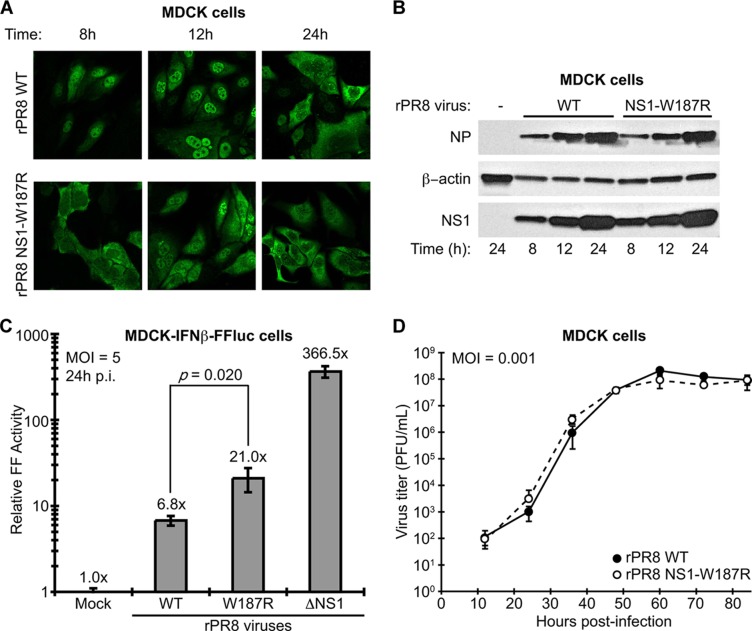
Characterization of WT and NS1-W187R viruses in vitro. (A) NS1 localization during infection. Shown is indirect immunofluorescence analysis of NS1 protein localization in MDCK cells infected for the indicated times with rPR8 WT or rPR8 NS1-W187R viruses (multiplicity of infection [MOI] of 2 PFU/cell). The primary antibody was pAb 155 (12). (B) Expression of viral NS1 and NP proteins during infection. SDS-PAGE and Western blot analysis of lysates from MDCK cells infected for the indicated times with rPR8 WT or rPR8 NS1-W187R viruses (MOI of 5 PFU/cell). NS1 was detected using pAb 155, NP was detected using monoclonal antibody (MAb) HT103 (20), and β-actin was detected using MAb A4700 (Sigma-Aldrich, St. Louis, MO). (C) Induction of IFN-β by different rPR8 mutants. MDCK-IFN-β-FF-Luc cells were infected at an MOI of 5 PFU/cell for 24 h with the indicated virus (or mock infected) prior to analysis of luciferase activity. p.i., postinfection. Bars represent mean values (n = 3), and error bars represent standard deviations (SD). (D) Multicycle growth analysis of rPR8 WT and rPR8 NS1-W187R viruses in MDCK cells. Data points show mean values (n = 3), and error bars represent SD.

Characterization of WT and NS1-W187R virus replication and pathogenicity in vivo. Six- to 8-week-old C57/BL6 mice were infected intranasally with 32 PFU of each virus (10 mice per group). Body weights were determined daily, and mice showing more than 25% weight loss were considered to have reached the experimental endpoint and were humanely euthanized. (A) Survival data; (B) mean body weights. Error bars represent SD. Statistical significance (**, P < 0.0005) was determined using Student's t test. (C) Six- to 8-week-old C57/BL6 mice were infected intranasally with 1,250 PFU of each virus. Lung titers were determined on days 2 and 4 postinfection from 3 mice per group. Bars represent mean values. P values were determined using Student's t test.
References
-
- Cheng A, Wong SM, Yuan YA. 2009. Structural basis for dsRNA recognition by NS1 protein of influenza A virus. Cell Res. 19:187–195 - PubMed
-
- DeLano WL. 2002. The PyMOL molecular graphics system. DeLano Scientific, Palo Alto, CA
Publication types
MeSH terms
Substances
Grants and funding
LinkOut - more resources
Full Text Sources
