. 2013 Jan 4;112(1):48-56.
doi: 10.1161/CIRCRESAHA.112.272963. Epub 2012 Sep 19.

Unrestrained p38 MAPK activation in Dusp1/4 double-null mice induces cardiomyopathy

Affiliations

Unrestrained p38 MAPK activation in Dusp1/4 double-null mice induces cardiomyopathy

Mannix Auger-Messier et al. Circ Res. .

Erratum in

  • Circ Res. 2013 Feb 1;112(3):e32

Abstract

Rationale: Mitogen-activated protein kinases (MAPKs) are activated in the heart by disease-inducing and stress-inducing stimuli, where they participate in hypertrophy, remodeling, contractility, and heart failure. A family of dual-specificity phosphatases (DUSPs) directly inactivates each of the MAPK terminal effectors, potentially serving a cardioprotective role.

Objective: To determine the role of DUSP1 and DUSP4 in regulating p38 MAPK function in the heart and the effect on disease.

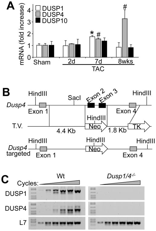
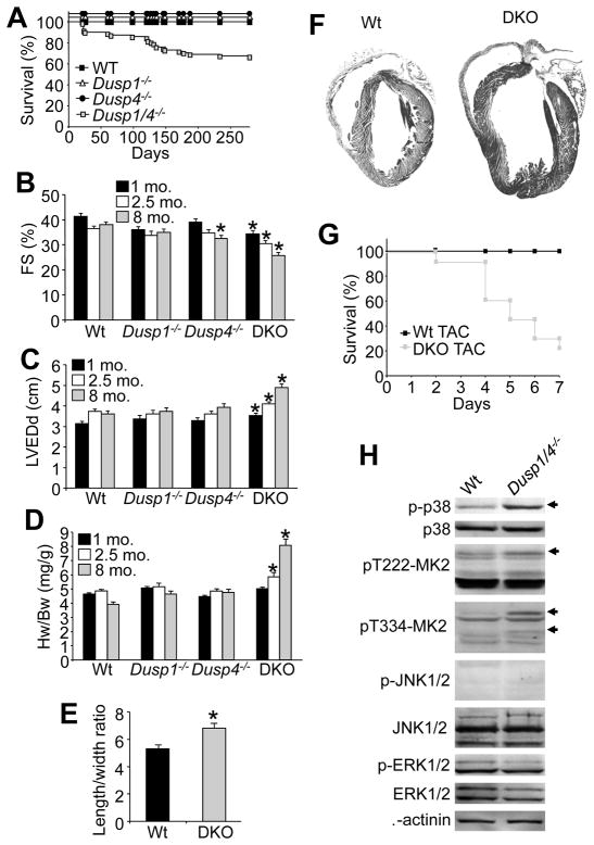
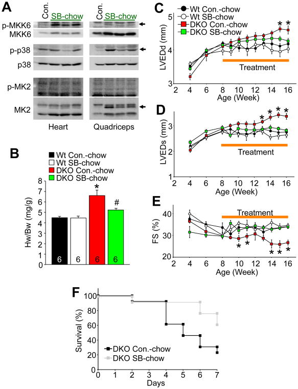
Methods and results: Here, we generated mice and mouse embryonic fibroblasts lacking both Dusp1 and Dusp4 genes. Although single nulls showed no molecular effects, combined disruption of Dusp1/4 promoted unrestrained p38 MAPK activity in both mouse embryonic fibroblasts and the heart, with no change in the phosphorylation of c-Jun N-terminal kinases or extracellular signal-regulated kinases at baseline or with stress stimulation. Single disruption of either Dusp1 or Dusp4 did not result in cardiac pathology, although Dusp1/4 double-null mice exhibited cardiomyopathy and increased mortality with aging. Pharmacological inhibition of p38 MAPK with SB731445 ameliorated cardiomyopathy in Dusp1/4 double-null mice, indicating that DUSP1/4 function primarily through p38 MAPK in affecting disease. At the cellular level, unrestrained p38 MAPK activity diminished cardiac contractility and Ca2+ handling, which was acutely reversed with a p38 inhibitory compound. Poor function in Dusp1/4 double-null mice also was partially rescued by phospholamban deletion.

Conclusions: Our data demonstrate that Dusp1 and Dusp4 are cardioprotective genes that play a critical role in the heart by dampening p38 MAPK signaling that would otherwise reduce contractility and induce cardiomyopathy.


Figures

Figure 1
Figure 1. Gene-targeting of stress-responsive Dusp4 in the heart
A, Normalized SYBR green quantitative PCR for DUSP1, DUSP4, and DUSP10 encoding mRNA from heart of mice subjected to Sham or TAC procedure for the indicated period of time in days or weeks (*P < 0.01 vs. sham; #P < 0.05 vs. sham; n=3–6 per time point). B, Schematic of the Dusp4 genetic locus and the targeting vector (T.V.) used to create Dusp4 gene-deleted mice. C, RT-PCR for DUSP1, DUSP4, and L7 (control) from hearts of control (Wt) and Dusp1/4 double null (Dusp1/4−/−) mice at 1 month of age.
Figure 2
Figure 2. Combined deletion of Dusp1 and Dusp4 results in unrestrained p38 MAPK phosphorylation
A, Western blot analysis of key MAPK pathway enzymes from serum-cultured MEFs derived from Wt or Dusp1/4−/− embryos. Arrowheads indicate changed band intensities detected from Dusp1/4−/− cells. B, Western blot analysis of MAPK phosphorylation from Wt or Dusp1/4−/− MEFs stimulated with anisomycin (1 μg/ml) for the indicated period of time under serum-starved conditions. Asterisk shows expanded p38 phosphorylation. C, Western blot analysis of the indicated proteins and phospho-proteins from hearts of adult mice subjected to sham or TAC procedure (15 minutes of stimulation) in Wt or Dusp1/4−/− mice previously fed vehicle or SB731445-formulated chow. Asterisks and arrow heads in the gel picture or bottom of it show bands that are differentially regulated by loss of Dusp1/4, while the 2 arrowheads on the right side show the migration of MK2. D, Western blot analysis of phosphorylated and total p38 MAPK from the hearts of 2 week-old Wt or Dusp1/4−/− mice injected with anisomycin systemically (10 mg/kg) for the indicated period of time. Asterisks show expanded p38 phosphorylation. E, Western blot analysis of phosphorylated and total p38 MAPK from the hearts of 2 month-old Wt or Dusp1/4−/− mice subjected to Sham or TAC procedure for the indicated periods of time in minutes or hours.
Figure 3
Figure 3. Dusp1/4−/− mice spontaneously develop cardiomyopathy with aging
A, Kaplan-Meier survival curve of Wt (black square, n=10), Dusp1−/− (triangle, n=10), Dusp4−/− (circle, n=10), and Dusp1/4−/− (grey square, n=71) mice. (*p<0.01 vs. other genotypes). B and C, Assessment of fractional shortening (FS) and left ventricular end dimension in diastole (LVEDd) by echocardiography in Wt (n=8), Dusp1−/− (n=8), Dusp4−/− (n=8), and Dusp1/4−/− (DKO, n=17) mice at 1, 2, and 8 months of age. (*p<0.01 vs. Wt). D, Heart weight (Hw) normalized to body weight (Bw) in Wt, Dusp1−/−, Dusp4−/−, and Dusp1/4−/− (DKO) mice at 1, 2, and 8 months of age (n=10 per genotype/age). (*p<0.01 vs. Wt). E, Length/width ratio from Wt and Dusp1/4−/− (DKO) isolated adult cardiomyocytes (*p<0.01 vs. Wt; n=200 cells/genotype). F, Gross H&E-stained histological section in longitudinal section through hearts of the indicated mice at 8 months of age. G, Kaplan-Meier survival curve over 7 days after TAC stimulation in adult Wt or DKO mice (N=12 or greater mice per group at onset). H, Western blot analysis of MAPK pathway enzymes from Wt or Dusp1/4−/− isolated adult cardiomyocytes cultured in the absence of serum for 16h. Arrows indicate increased phospho-specific bands detected from Dusp1/4−/− cells.
Figure 4
Figure 4. Pharmacological inhibition of p38 MAPK prevents cardiomyopathy in Dusp1/4−/− mice
A, Western blot analysis of key p38 MAPK pathway enzymes from heart or quadriceps of 2 month-old Wt mice fed with control (Con.) or p38 inhibitor SB731445-formulated chow for 1 week (50 mg/kg/day; SB-chow). Arrowheads indicate accelerated migration of MK2 characteristic of its unphosphorylated form, as well as the increase in MKK6 and phospho-p38. B, Heart weight (Hw) normalized to body weight (Bw) in 16 week-old Wt and Dusp1/4−/− (DKO) mice fed with either control chow (Con.-chow) or SB-chow from 8 to 16 weeks of age (*p<0.01 vs. Wt Con.-chow; #p<0.01 vs. DKO Con.-chow). The number of animals is indicated in the bars. C–E, Assessment of left ventricular end dimension in diastole (LVEDd), left ventricular end dimension in systole (LVEDs), and fractional shortening (FS) by echocardiography in Wt and Dusp1/4−/− (DKO) mice fed with either control chow (Con.-chow) or SB-chow from 8 to 16 weeks of age, although echo assessment began 4 weeks prior to treatment (*p<0.01 vs. other genotypes). F, Kaplan-Meier survival curve over 7 days after TAC stimulation in adult DKO mice with prior and continuous vehicle or SB731445-containing chow treatment (N=12 or greater mice per group at onset).
Figure 5
Figure 5. Inhibition of p38 MAPK restores depressed contractility in Dusp1/4−/− mice and isolated cardiomyocytes
A and B, Invasive hemodynamic assessment of contraction (Max dP/dt) and relaxation (Min dP/dt) velocities in anesthetized, close-chested Wt and Dusp1/4−/− (DKO) mice fed for 2 weeks with either control chow (Con.-chow) or p38 inhibitor SB731445-formulated chow (50 mg/kg/day; SB-chow). (N=5 or more mice per group) (*p<0.01 vs. Wt Con.-chow; #p<0.01 vs. DKO Con.-chow). C, Representative traces of fura-2 F340/F380 fluorescence ratio (black trace) and myocytes shortening (red trace) recordings from Wt or Dusp1/4−/− (DKO) isolated adult cardiomyocytes in the absence or presence of p38 inhibitor SB239063 (50 μM; SB). D–G, Average maximal peak amplitude of electrically evoked Ca2+ transients, time constant of Ca2+ decay, average maximal Ca2+ response to a 10 mM caffeine bolus, and percentage of shortening in isolated adult cardiomyocytes from the indicated genotypes in absence (Control) or presence of p38 inhibitor SB239063 (50 μM; SB). At least 4 animals were used, and the total number of cells analyzed is indicated in the bars (*p<0.05 vs. Wt Control; #p<0.05 vs. DKO Control).
Figure 6
Figure 6. Deletion of Pln prevents cardiomyopathy in Dusp1/4 DKO mice
A, Assessment of fractional shortening (FS %), B, left ventricular end dimension in diastole (LVEDd), and C, left ventricular end dimension in systole (LVEDs) in the indicated groups of mice at 4 months of age. Number of mice analyzed is shown in the bars. *P<0.05 versus Wt; #P<0.05 versus Dusp1/4 DKO mice

References

    1. Garrington TP, Johnson GL. Organization and regulation of mitogen-activated protein kinase signaling pathways. Curr Opin Cell Biol. 1999;11:211–218. - PubMed
    1. Baines CP, Molkentin JD. STRESS signaling pathways that modulate cardiac myocyte apoptosis. J Mol Cell Cardiol. 2005;38:47–62. - PubMed
    1. Rose BA, Force T, Wang Y. Mitogen-activated protein kinase signaling in the heart: angels versus demons in a heart-breaking tale. Physiol Rev. 2010;90:1507–1546. - PMC - PubMed
    1. Liao P, Georgakopoulos D, Kovacs A, Zheng M, Lerner D, Pu H, Saffitz J, Chien K, Xiao RP, Kass DA, Wang Y. The in vivo role of p38 MAP kinases in cardiac remodeling and restrictive cardiomyopathy. Proc Natl Acad Sci USA. 2001;98:12283–12288. - PMC - PubMed
    1. Nishida K, Yamaguchi O, Hirotani S, Hikoso S, Higuchi Y, Watanabe T, Takeda T, Osuka S, Morita T, Kondoh G, Uno Y, Kashiwase K, Taniike M, Nakai A, Matsumura Y, Miyazaki J-I, Sudo T, Hongo K, Kusakari Y, Kurihara S, Chien KR, Takeda J, Hori M, Otsu K. p38α mitogen-activated protein kinase plays a critical role in cardiomyocyte survival but not in cardiac hypertrophic growth in response to pressure overload. Mol Cell Biol. 2004;24:10611–10620. - PMC - PubMed

Publication types

MeSH terms