. 2012 Sep 19;32(38):13100-10.
doi: 10.1523/JNEUROSCI.2604-12.2012.

Astrocyte-derived thrombospondins mediate the development of hippocampal presynaptic plasticity in vitro

Affiliations

Astrocyte-derived thrombospondins mediate the development of hippocampal presynaptic plasticity in vitro

Devon C Crawford et al. J Neurosci. .

Abstract

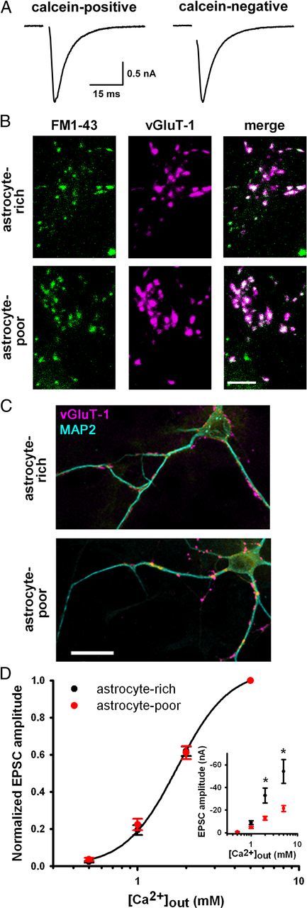
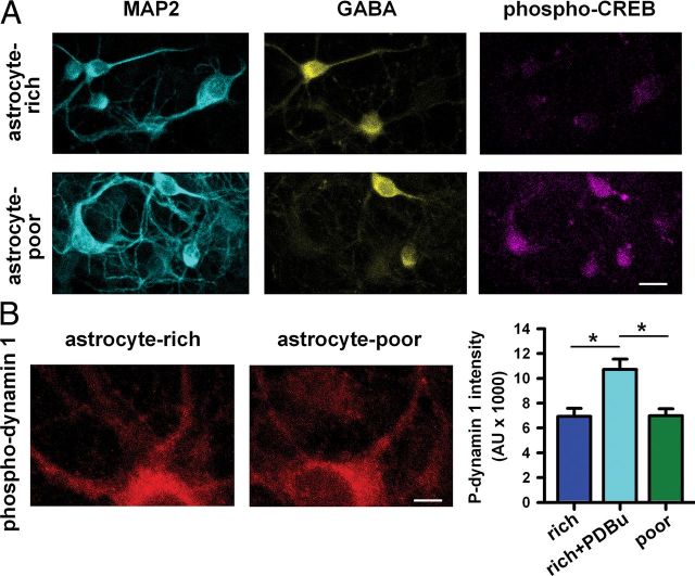
Astrocytes contribute to many neuronal functions, including synaptogenesis, but their role in the development of synaptic plasticity remains unclear. Presynaptic muting of hippocampal glutamatergic terminals defends against excitotoxicity. Here we studied the role of astrocytes in the development of presynaptic muting at glutamatergic synapses in rat hippocampal neurons. We found that astrocytes were critical for the development of depolarization-dependent and G(i/o)-dependent presynaptic muting. The ability of cAMP analogues to modulate presynaptic function was also impaired by astrocyte deficiency. Although astrocyte deprivation resulted in postsynaptic glutamate receptor deficits, this effect appeared independent of astrocytes' role in presynaptic muting. Muting was restored with chronic, but not acute, treatment with astrocyte-conditioned medium, indicating that a soluble factor is permissive for muting. Astrocyte-derived thrombospondins (TSPs) are likely responsible because TSP1 mimicked the effect of conditioned medium, and gabapentin, a high-affinity antagonist of TSP binding to the α2δ-1 calcium channel subunit, mimicked astrocyte deprivation. We found evidence that protein kinase A activity is abnormal in astrocyte-deprived neurons but restored by TSP1, so protein kinase A dysfunction may provide a mechanism by which muting is disrupted during astrocyte deficiency. In summary our results suggest an important role for astrocyte-derived TSPs, acting through α2δ-1, in maturation of a potentially important form of presynaptic plasticity.


Figures

Figure 1.
Figure 1.
Astrocyte deprivation impairs presynaptic muting but not G-protein activation. A, Phase-contrast and fluorescence images of autaptic neurons on viable (control) or nonviable (4% paraformaldehyde/0.2% glutaraldehyde-fixed) glial “islands” acquired 30 min after 5 μg/ml calcein-AM treatment. Fewer islands contained live (calcein-positive) astrocytes in fixed cultures (control: 98.8 ± 0.9% live islands; fixed: 17.6 ± 2.8% live islands; n = 6 dishes; p = 7.9 × 10⁻¹¹, Student's unpaired t test). Scale bar, 40 μm. B, Representative autaptic EPSCs from astrocyte-rich or astrocyte-poor microcultures recorded in normal saline up to 1 h after 4 h control treatment (30 mm NaCl) or after 4 h depolarization (30 mm KCl). C, Summary of peak EPSC amplitudes from DIV 10–14 astrocyte-rich (+) or astrocyte-poor (−) autaptic neurons after indicated treatment times with 30 mm KCl (black bars), 10 nm CCPA (A1 adenosine receptor agonist) plus 50 μm baclofen (GABAB receptor agonist) (C+B; yellow bars), or 50 μm Rp-cAMPS (Rp; cyan bars). EPSCs were normalized to the average EPSCs from control-treated sibling neurons for each recording day (n = 14–22 neurons; *p < 0.05 vs treatment controls, Student's unpaired t test). D, EPSCs from astrocyte-rich or astrocyte-poor cultures after acute perfusion with recording saline (control) or 20 μm baclofen. E, EPSCs from astrocyte-rich or astrocyte-poor cultures after acute perfusion with recording saline (control) or 10 nm CCPA. The same neurons were used for experiments in D and E. D, E, Summary data are given in the Results.
Figure 2.
Figure 2.
Astrocyte deprivation does not impair basal presynaptic function. A, EPSCs from autaptic neurons in astrocyte-poor cultures from islands with residual live astrocytes (calcein-positive) or no live astrocytes (calcein-negative). B, Images of 10 μm FM1-43FX labeling of presynaptic terminals (green) after 2 min 45 mm KCl and subsequent vGluT-1 immunostaining (magenta) in autaptic neurons from astrocyte-rich and astrocyte-poor cultures (30 min, −20°C, 70% EtOH-fixed astrocytes). Scale bar, 5 μm. C, vGluT-1 (magenta) and MAP2 (cyan) immunostaining in autaptic neurons showing similar density and intensity of presynaptic terminals. Scale bar, 20 μm. Summary results for A–C are given in the Results. D, Calcium concentration–response curves of autaptic EPSC amplitudes from astrocyte-rich or astrocyte-poor cultures normalized for each neuron to values obtained in 5 mm external calcium (n = 21–22). Data from astrocyte-rich cultures were fitted to the Hill equation (EC50 value: 1.8 mm; Hill coefficient: 2.7). Inset, Non-normalized values from the same dataset demonstrating basal depression of EPSCs in astrocyte-poor cultures at higher calcium concentrations. *p < 0.05, Student's unpaired t test, Bonferroni corrected.
Figure 3.
Figure 3.
Glutamate receptor levels are decreased in astrocyte-poor cultures. A, Representative dual-component (NMDA and AMPA receptor-mediated) EPSCs elicited in autaptic neurons from astrocyte-rich or astrocyte-poor cultures. Note the change in vertical scale bar. B, Top, Currents from autaptic neurons in astrocyte-rich or astrocyte-poor cultures in response to acute kainic acid (KA) perfusion, a nondesensitizing AMPA receptor agonist. Bottom, Currents from autaptic neurons in astrocyte-rich or astrocyte-poor cultures in response to acute NMDA perfusion to selectively activate NMDA receptors. C, Top, representative miniature EPSCs from astrocyte-rich or astrocyte-poor cultures elicited by acute perfusion of 100 mm sucrose. Bottom, Cumulative probability plots of amplitude (p < 0.001 with D = 0.2175) and interevent interval (p < 0.001 with D = 0.3860) of miniature EPSCs (n = 10; Kolmogorov–Smirnov test). Additional summary data for A–C are given in the Results.
Figure 4.
Figure 4.
Soluble, astrocyte-derived thrombospondin rescues presynaptic muting competence. A–D, Representative DIV 7–12 autaptic EPSCs after 4 h control treatment (30 mm NaCl) or depolarization (30 mm KCl). Cultures were astrocyte-rich and challenged at DIV 7 without additional treatments (A), astrocyte-poor, treated with astrocyte-conditioned medium (CM) at DIV 7, and challenged at DIV 10–12 (B), astrocyte-poor and challenged at DIV 10–12 in CM (C), or astrocyte-poor, treated with 5 μg/ml human thrombospondin 1 (TSP) at DIV 7, and challenged at DIV 10–12 (D). E, Summary of EPSC amplitudes after 4 h 30 mm KCl or 50 μm Rp-cAMPS (Rp) with indicated pretreatments and/or cotreatments (n = 12–20 neurons; *p < 0.05 vs treatment controls, Student's unpaired t test). EPSCs were normalized as in Figure 1C. +, Astrocyte-rich cultures; −, astrocyte-poor cultures; GBP, 32 μM gabapentin; TNFα, 1 ng/ml tumor necrosis factor α; FM, fresh, non-conditioned medium.
Figure 5.
Figure 5.
Gabapentin prevents the development of muting competence. A, Representative DIV 10–12 autaptic EPSCs after 4 h control treatment (30 mm NaCl) or depolarized (30 mm KCl). Astrocyte-rich cultures were treated with 32 μm gabapentin at DIV 7 (1× GBP). B, Representative DIV 10–12 autaptic EPSCs after 4 h control or depolarized. Astrocyte-rich cultures were treated with 32 μm gabapentin at DIV 7 followed by half-media switches of fresh 32 μm gabapentin in astrocyte-conditioned medium every day (3–5× GBP). Summary data for A and B are given in the Results. C, Spontaneous activity in DIV 11–13 mass cultures after 4 h control treatment (30 mm NaCl) or depolarized (30 mm KCl). Cultures were treated at DIV 7 with or without 32 μm gabapentin followed by daily half media switches with astrocyte-conditioned medium (CM) or CM plus 32 μm gabapentin (3–5× GBP). EPSCs and IPSCs were measured simultaneously at −35 mV. D, Summary of total spontaneous synaptic activity (EPSC + IPSC points) in DIV 11–13 mass cultures, normalized to control (CM, NaCl) for each recording day (n = 23 neurons; *p < 0.05, Bonferroni corrected).
Figure 6.
Figure 6.
PKA-dependent hyperphosphorylation of synapsin is normalized by thrombospondin. A, Phospho-synapsin (PKA phosphorylation site) immunostaining in DIV 10–12 autaptic neurons from astrocyte-rich or astrocyte-poor cultures with or without 5 μg/ml TSP treatment at DIV 7. Red circles are representative regions of interest defined by vGluT-1 immunoreactivity (data not shown). Scale bar, 5 μm. B, Summary of background-subtracted phospho-synapsin at vGluT-1-positive synapses in DIV 10–12 autaptic neurons (n = 35 neurons; *p < 0.05, Bonferroni corrected). C, Synapsin immunostaining at vGluT-1-defined synapses (red circles are representative) in autaptic neurons from astrocyte-rich or astrocyte-poor cultures. Summary immunoreactivity values are given in the Results. Scale bar, 5 μm.
Figure 7.
Figure 7.
A PKA target, but not a non-PKA target, is hyperphosphorylated in astrocyte-poor cultures. A, MAP2, GABA, and phospho-CREB (PKA phosphorylation site) immunoreactivity in neurons on multicell islands from astrocyte-rich or astrocyte-poor cultures. Summary pCREB immunoreactivity values are given in the Results. Scale bar, 20 μm. B, Left, Phospho-dynamin 1 (PKC phosphorylation site) immunoreactivity in autaptic neurons from astrocyte-rich and astrocyte-poor cultures. Scale bar, 20 μm. Right, Quantification of background-subtracted phospho-dynamin 1 intensity from a single primary and two secondary dendrites averaged per neuron. As a positive control, we treated for 30 min with phorbol ester (1 μm PDBu), which increased PKC-dependent phosphorylation of dynamin 1 (n = 30 neurons; *p < 0.05, Bonferroni corrected).

References

    1. Adams JC, Tucker RP. The thrombospondin type 1 repeat (TSR) superfamily: diverse proteins with related roles in neuronal development. Dev Dyn. 2000;218:280–299. - PubMed
    1. Alexanian AR. Neural stem cells induce bone-marrow-derived mesenchymal stem cells to generate neural stem-like cells via juxtacrine and paracrine interactions. Exp Cell Res. 2005;310:383–391. - PubMed
    1. Amur-Umarjee S, Phan T, Campagnoni AT. Myelin basic protein mRNA translocation in oligodendrocytes is inhibited by astrocytes in vitro. J Neurosci Res. 1993;36:99–110. - PubMed
    1. Arikkath J, Campbell KP. Auxiliary subunits: essential components of the voltage-gated calcium channel complex. Curr Opin Neurobiol. 2003;13:298–307. - PubMed
    1. Asch AS, Leung LL, Shapiro J, Nachman RL. Human brain glial cells synthesize thrombospondin. Proc Natl Acad Sci U S A. 1986;83:2904–2908. - PMC - PubMed

Publication types

MeSH terms

Substances

LinkOut - more resources