. 2012 Sep 19;32(38):13155-63.
doi: 10.1523/JNEUROSCI.2530-12.2012.

Peripheral ammonia as a mediator of methamphetamine neurotoxicity

Affiliations

Peripheral ammonia as a mediator of methamphetamine neurotoxicity

Laura E Halpin et al. J Neurosci. .

Abstract

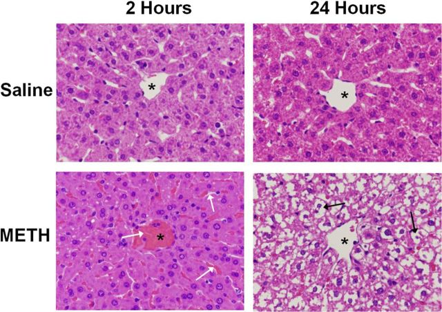
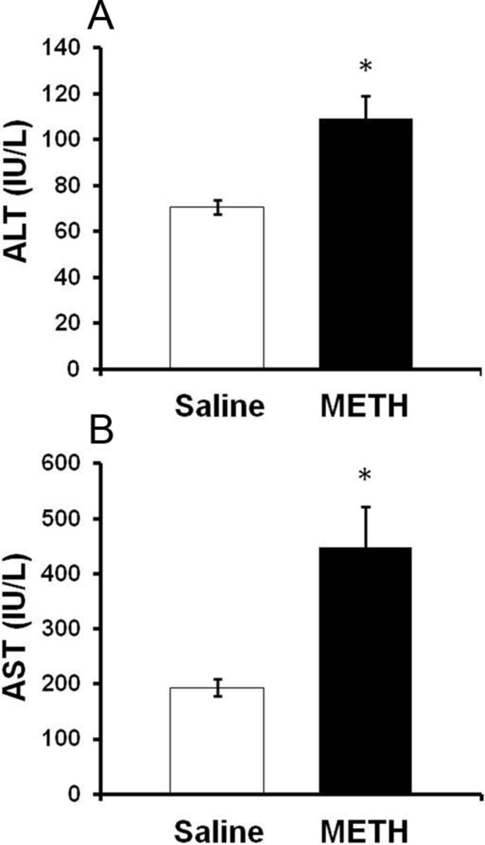
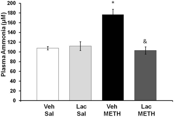
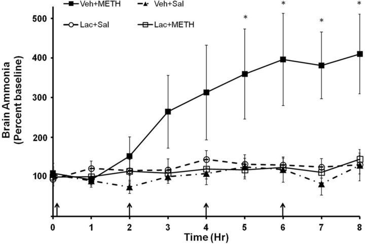
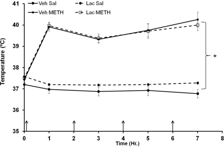
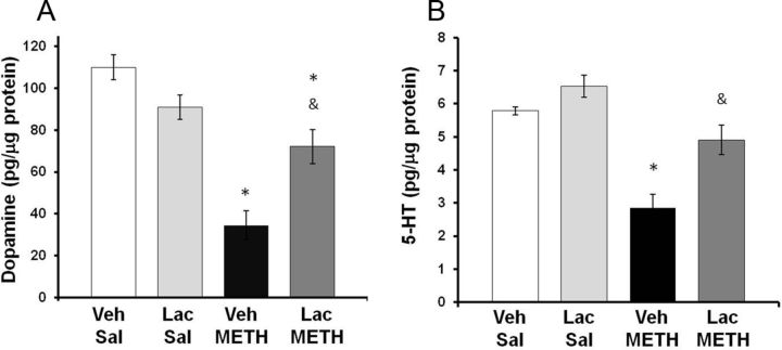
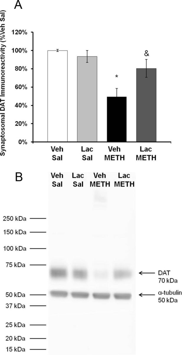
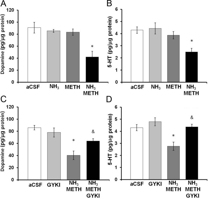
Ammonia is metabolized by the liver and has established neurological effects. The current study examined the possibility that ammonia contributes to the neurotoxic effects of methamphetamine (METH). The results show that a binge dosing regimen of METH to the rat increased plasma and brain ammonia concentrations that were paralleled by evidence of hepatotoxicity. The role of peripheral ammonia in the neurotoxic effects of METH was further substantiated by the demonstration that the enhancement of peripheral ammonia excretion blocked the increases in brain and plasma ammonia and attenuated the long-term depletions of dopamine and serotonin typically produced by METH. Conversely, the localized perfusion of ammonia in combination with METH, but not METH alone or ammonia alone, into the striatum recapitulated the neuronal damage produced by the systemic administration of METH. Furthermore, this damage produced by the local administration of ammonia and METH was blocked by the GYKI 52466 [4-(8-methyl-9H-1,3-dioxolo[4,5-h][2,3]benzodiazepin-5-yl)-benzamine hydrochloride], an AMPA receptor antagonist. These findings highlight the importance of ammonia derived from the periphery as a small-molecule mediator of METH neurotoxicity and more broadly emphasize the importance of peripheral organ damage as a possible mechanism that mediates the neuropathology produced by drugs of abuse and other neuroactive molecules.


Figures

Figure 1.
Figure 1.
METH causes hepatocellular damage visible by hematoxylin and eosin staining. Hematoxylin and eosin staining was used to examine the effects of METH (10 mg/kg, i.p., every 2 h × 4) or saline (1 ml/kg, i.p., every 2 h × 4) on the livers of treated rats. Livers were examined at 2 and 24 h after the last injection of METH or saline. A representative image of a hepatic lobule near the central vein is shown (asterisks denote central vein). The images in the figure are representative of all rats examined for each treatment and at each time point. At 2 h, there is moderate vascular and sinusoidal congestion evident throughout the livers of METH-treated rats (white arrows). At 24 h, this congestion leads to extensive cytoplasmic disappearance in the livers of all METH-treated rats (black arrows) (magnification, 40×).
Figure 2.
Figure 2.
Serum AST and ALT after METH treatment. A, B, Rats were treated with METH (10 mg/kg, i.p., every 2 h × 4) or saline (1 ml/kg, i.p., every 2 h × 4). At 2 h after the last injection of METH or saline, (A) ALT and (B) AST levels were significantly elevated to 54.9 ± 15.2% and 141.0 ± 44.1% in METH-treated rats compared with saline controls, respectively. Values are mean ± SEM, *p < 0.05 (n = 12 rats per group).
Figure 3.
Figure 3.
METH and plasma ammonia. Rats were treated with METH (10 mg/kg, i.p., every 2 h × 4) or saline (1 ml/kg, i.p., every 2 h × 4) and lactulose (5.3 g/kg, p.o., every 12 h) or vehicle (8 ml/kg, p.o., every 12 h). At 2 h after the last injection of METH or saline, plasma ammonia levels were significantly elevated by 64.04 ± 9.98% (mean ± SEM) and this increase was blocked by lactulose treatment (*p < 0.001 compared with Lac Sal, &p < 0.001 compared with Veh METH) (n = 8 rats per group). Lac, lactulose; Sal, saline; Veh, vehicle.
Figure 4.
Figure 4.
METH and brain ammonia. Rats were treated with METH (10 mg/kg × 4, i.p., every 2 h) or saline (1 ml/kg × 4, i.p., every 2 h) and lactulose (5.3 g/kg, p.o., every 12 h) or vehicle (8 ml/kg, p.o., every 12 h). During drug treatment (arrows denote injections), striatal ammonia concentration was measured using in vivo microdialysis. Vehicle plus METH treatment significantly increased brain ammonia concentrations starting at time point 5 and lactulose pretreatment attenuated this increase (*p < 0.05). Baseline, 29.34 ± 5.33 μm. (n = 8–10 rats per group). Lac, lactulose; Sal, saline; Veh, vehicle.
Figure 5.
Figure 5.
Hyperthermia after METH treatment. Rats were treated with METH (10 mg/kg, i.p., every 2 h × 4) or saline (1 ml/kg, i.p., every 2 h × 4) and lactulose (5.3 g/kg, p.o., every 12 h) or vehicle (8 ml/kg, p.o., every 12 h). During drug treatment, temperature was measured 1 h after each METH injection (denoted by arrows). METH treatment caused significant hyperthermia in both vehicle plus METH-treated and lactulose plus METH-treated rats, and lactulose had no significant effect on hyperthermia (*p < 0.001 compared with Veh Sal) (n = 6–9 per group). Lac, lactulose; Sal, saline; Veh, vehicle.
Figure 6.
Figure 6.
Effect of lactulose on METH-induced monoamine depletions. Rats were treated with METH (10 mg/kg, i.p., every 2 h × 4) or saline (1 ml/kg, i.p., every 2 h × 4) and lactulose (5.3 g/kg, p.o., every 12 h) or vehicle (8 ml/kg, p.o., every 12 h). Tissue content of dopamine or 5-HT in the striatum was measured 7 d later. A, Treatment with lactulose significantly attenuated the striatal dopamine depletions caused by METH as METH depleted dopamine by only 34.5 ± 7.9% (mean ± SEM) in lactulose-treated rats. B, METH depleted 5-HT by 15.2 ± 8.2% (mean ± SEM) in lactulose-treated rats, which was not statistically significant compared with saline-treated rats (*p < 0.001 compared with Lac Sal; &p < 0.001 compared with Veh METH) (n = 6–9 rats per group). Lac, lactulose; Sal, saline; Veh, vehicle.
Figure 7.
Figure 7.
Effect of lactulose on METH-induced reductions in DAT immunoreactivity. Rats were treated with METH (10 mg/kg, i.p., every 2 h × 4) or saline (1 ml/kg, i.p., every 2 h × 4) and lactulose (5.3 g/kg, p.o., every 12 h) or vehicle (8 ml/kg, p.o., every 12 h). Tissue content of dopamine or 5-HT in the striatum were measured 7 d later. A, Treatment with lactulose significantly blocked the decrease in DAT immunoreactivity as METH produced only a 19.5 ± 9.8% depletion in lactulose plus METH-treated rats. B, Representative Western blot for DAT immunoreactivity and α-tubulin internal control (*p < 0.05 compared with Lac Sal. &p < 0.05 compared with Veh METH) (n = 6 rats per group). Lac, lactulose; Sal, saline; Veh, vehicle.
Figure 8.
Figure 8.
Effects of local administration of ammonia, METH, and GYKI 52466 into the striatum. Rats were reverse dialyzed with METH (100 μm), ammonia (570 μm), and/or GKYI 52466 (100 μm) in the striatum for 8 h. Dopamine and 5-HT tissue content of the striatal tissue around the probe membrane (1 mm) was measured 7 d later. A, Treatment with the combination of METH and ammonia depletes dopamine in the tissue around the probe by 51.5 ± 12.5% (mean ± SEM). B, Treatment with the combination of METH and ammonia depleted 5-HT in the tissue around the probe by 41.8 ± 7.0% (mean ± SEM) compared with aCSF. C, Treatment with GYKI 52466 significantly attenuated depletions in dopamine tissue content produced by METH and ammonia. D, Treatment with GKYI 52466 significantly attenuated depletions in 5-HT tissue content produced by the combination of METH and ammonia (*p < 0.05 compared with aCSF, &p < 0.05 compared with ammonia METH) (n = 10 rats per group). GYKI, GYKI 52466.

Similar articles

Cited by

References

    1. Ago M, Ago K, Hara K, Kashimura S, Ogata M. Toxicological and histopathological analysis of a patient who died nine days after a single intravenous dose of methamphetamine: a case report. Leg Med (Tokyo) 2006;8:235–239. - PubMed
    1. Al Sibae MR, McGuire BM. Current trends in the treatment of hepatic encephalopathy. Ther Clin Risk Manag. 2009;5:617–626. - PMC - PubMed
    1. Batshaw ML, Hyman SL, Mellits ED, Thomas GH, DeMuro R, Coyle JT. Behavioral and neurotransmitter changes in the urease-infused rat: a model of congenital hyperammonemia. Pediatr Res. 1986;20:1310–1315. - PubMed
    1. Breier JM, Bankson MG, Yamamoto BK. l-tyrosine contributes to (+)-3,4-methylenedioxymethamphetamine-induced serotonin depletions. J Neurosci. 2006;26:290–299. - PMC - PubMed
    1. Brown JM, Riddle EL, Sandoval V, Weston RK, Hanson JE, Crosby MJ, Ugarte YV, Gibb JW, Hanson GR, Fleckenstein AE. A single methamphetamine administration rapidly decreases vesicular dopamine uptake. J Pharmacol Exp Ther. 2002;302:497–501. - PubMed

Publication types

MeSH terms