. 2010 Sep;1(5):809-816.
doi: 10.3892/etm.2010.132. Epub 2010 Jul 21.

Characteristic expression pattern of oxidative stress in livers with cryptogenic hepatocellular carcinoma

Affiliations

Characteristic expression pattern of oxidative stress in livers with cryptogenic hepatocellular carcinoma

Nobuyoshi Fukushima et al. Exp Ther Med. 2010 Sep.

Abstract

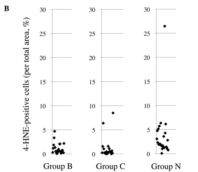
The mechanism responsible for the development of hepatocellular carcinoma (HCC) in the setting of oxidative stress has yet to be clearly defined. We studied the role of oxidative stress in hepatocarcinogenesis in subjects without underlying chronic viral hepatitis. The subjects were 24 patients negative for serum hepatitis B surface antigen and hepatitis C antibody tests, who underwent hepatic resection for HCC (Group N). Subjects were excluded if diagnosed with liver disease predisposing to HCC. Immunohistochemical staining for oxidative stress-related markers was performed on non-cancerous liver regions. Resected liver tissues adjacent to HCC from 24 patients with chronic hepatitis B (Group B) and 21 patients with chronic hepatitis C (Group C) were also examined. The percentage of 8-hydroxydeoxyguanosine-positive hepatocytes in Group N was significantly lower than that in Group B and that in the combined population of Groups B and C. The percentage of the area positive for 4-hydroxynonenal in Group N was significantly higher than that in Groups B or C. Meanwhile, the percentage of the area positive for manganese superoxide dismutase in Group N was not different from that in Groups B and C. In conclusion, the mechanism of hepatocarcinogenesis through oxidative stress for patients without known liver disease predisposing to HCC may differ from that for patients with chronic viral hepatitis.


Figures

Figure 1.
Figure 1.
Representative image of immunohistochemical staining in non-cancerous liver tissue from patients. (A) Staining for 8-OHdG in a patient with HCC negative for HCVAb and HBsAg (Group N). Many 8-OHdG-positive cells with a brown-stained nucleus were present in the section. Apart from hepatocytes, 8-OHdG-positive cells, such as Kupffer and infiltrative lymphocyte cells, were eliminated from the counting. Staining for 4-HNE (B) and Mn-SOD (C) in a patient with HCC negative for HCVAb and positive for HBsAg (Group B). Many 4-HNE- and Mn-SOD-positive cells were present in the section. Positive cells for both, except hepatocytes, were eliminated before quantitation.
Figure 1.
Figure 1.
Representative image of immunohistochemical staining in non-cancerous liver tissue from patients. (A) Staining for 8-OHdG in a patient with HCC negative for HCVAb and HBsAg (Group N). Many 8-OHdG-positive cells with a brown-stained nucleus were present in the section. Apart from hepatocytes, 8-OHdG-positive cells, such as Kupffer and infiltrative lymphocyte cells, were eliminated from the counting. Staining for 4-HNE (B) and Mn-SOD (C) in a patient with HCC negative for HCVAb and positive for HBsAg (Group B). Many 4-HNE- and Mn-SOD-positive cells were present in the section. Positive cells for both, except hepatocytes, were eliminated before quantitation.
Figure 2.
Figure 2.
Values for the ratios of the number of 8-OHdG-positive hepatocytes per 1,000 cells (A) and 4-HNE- (B) and Mn-SOD-positive cells (C) to the total area for each case in the three groups. Group N represents 24 patients with HCC negative for HCVAb and HBsAg, Group B represents 24 patients with HCC negative for HCVAb and positive for HBsAg, and Group C represents 21 patients with HCC positive for HCVAb, negative for HBsAg and antibody to hepatitis B core antigen in the serum.
Figure 2.
Figure 2.
Values for the ratios of the number of 8-OHdG-positive hepatocytes per 1,000 cells (A) and 4-HNE- (B) and Mn-SOD-positive cells (C) to the total area for each case in the three groups. Group N represents 24 patients with HCC negative for HCVAb and HBsAg, Group B represents 24 patients with HCC negative for HCVAb and positive for HBsAg, and Group C represents 21 patients with HCC positive for HCVAb, negative for HBsAg and antibody to hepatitis B core antigen in the serum.

Similar articles

References

    1. Wong F, Choi T. Primary liver cancer. Asian experience. In: Blumgart LH, editor. Surgery of Liver and Biliary Tract. Churchill-Livingstone; London: 1988. pp. 1135–1151.
    1. The Research Group for Population-based Cancer Resistration in Japan Cancer incidence and incidence rates in Japan in 1995: estimates based on data from nine population-based cancer registries. Jpn J Clin Oncol. 2000;30:318–321. - PubMed
    1. Tanaka H, Tsukuma H. Hepatitis C virus. In: Newton R, Beral V, Weiss RA, Tooze J, editors. Cancer Surveys: Infections and Human Cancer. Cold Spring Harbor Laboratory; New York: 1999. pp. 213–235.
    1. Di Bisceglie AM, Goodman ZD, Ishak KG, Hoofnagle JH, Melpolder JJ, Alter HJ. Long-term clinical and histopathological follow-up of chronic posttransfusion hepatitis. Hepatology. 1991;14:969–974. - PubMed
    1. Kiyosawa K, Sodeyama T, Tanaka E, et al. Interrelationship of blood transfusion, non-A, non-B hepatitis and hepatocellular carcinoma: analysis by detection of antibody to hepatitis C virus. Hepatology. 1990;12:671–675. - PubMed

LinkOut - more resources