. 2010 Sep;1(5):853-857.
doi: 10.3892/etm.2010.130. Epub 2010 Jul 21.

Gemcitabine and paclitaxel suppress the production of vascular endothelial growth factor induced by deferoxamine in human non-small cell lung cancer A549 cells

Affiliations

Gemcitabine and paclitaxel suppress the production of vascular endothelial growth factor induced by deferoxamine in human non-small cell lung cancer A549 cells

Ryuji Ikeda et al. Exp Ther Med. 2010 Sep.

Abstract

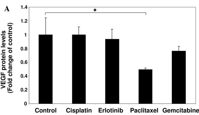
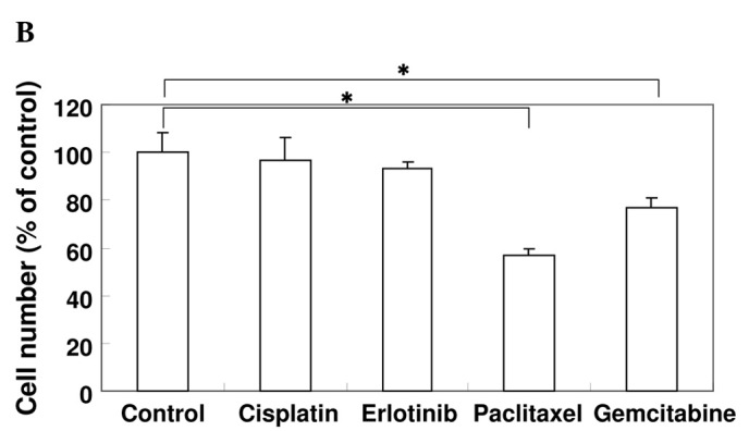
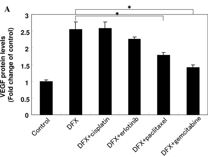
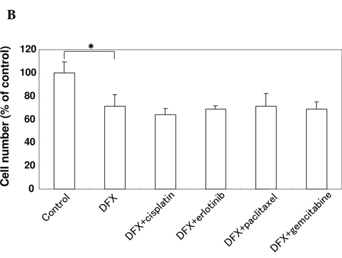
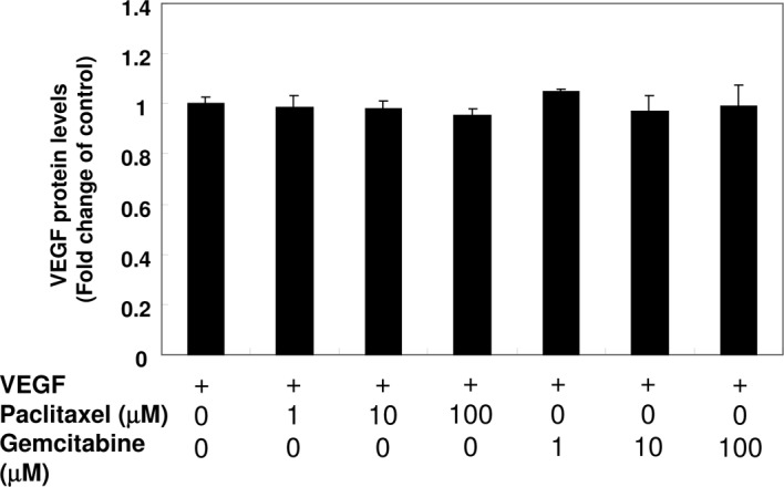
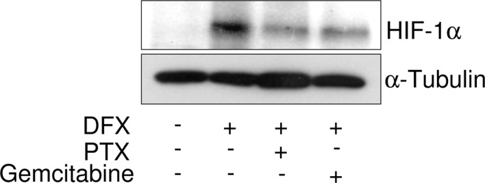
Vascular endothelial growth factor (VEGF) plays an important role in the process of angiogenesis in many types of cancer, including non-small cell lung cancer (NSCLC), and angiogenesis inhibitors and standard chemotherapy exhibit synergy though an unknown mechanism. We therefore hypothesized that cytotoxic chemotherapy influences VEGF production and analyzed VEGF production in an NSCLC A549 cell line after treatment with standard chemotherapy. Paclitaxel inhibited the production of VEGF in A549 cells, while cisplatin and erlotinib did not. Paclitaxel and gemcitabine inhibited deferoxamine (DFX) (known to mimic hypoxia)-induced VEGF production in A549 cells. Erlotinib also inhibited DFX-induced VEGF production in A549 cells slightly, while cisplatin did not. We subsequently examined the effect of the interaction between paclitaxel or gemcitabine and VEGF protein. Paclitaxel and gemcitabine did not directly affect the binding of VEGF. Since VEGF is known as one of the HIF-1 target genes, we examined the effect of paclitaxel and gemcitabine on HIF-1α levels induced by DFX in A549 cells. Paclitaxel and gemcitabine inhibited DFX-induced HIF-1α in A549 cells. These findings may be useful for future treatment schedules, including anti-cancer agents in NSCLC.


Figures

Figure 1.
Figure 1.
Effect of anti-cancer agents on the production of VEGF and cell proliferation. (A) A549 cells were cultured in serum-starved medium with or without 10 μM cisplatin, 10 μM erlotinib, 1 μM paclitaxel or 1 μM gemcitabine for 24 h as indicated. VEGF protein levels were measured using an ELISA assay. Data are the mean of triplicate experiments, and bars represent the mean ± SD. *P<0.05. (B) A549 cells were cultured in serum-starved medium with or without 10 μM cisplatin, 10 μM erlotinib, 1 μM paclitaxel or 1 μM gemcitabine for 24 h as indicated. The effect of the agents on cell proliferation was examined using the MTT assay. Data are the mean of triplicate experiments, and bars represent the mean ± SD. *P<0.05.
Figure 1.
Figure 1.
Effect of anti-cancer agents on the production of VEGF and cell proliferation. (A) A549 cells were cultured in serum-starved medium with or without 10 μM cisplatin, 10 μM erlotinib, 1 μM paclitaxel or 1 μM gemcitabine for 24 h as indicated. VEGF protein levels were measured using an ELISA assay. Data are the mean of triplicate experiments, and bars represent the mean ± SD. *P<0.05. (B) A549 cells were cultured in serum-starved medium with or without 10 μM cisplatin, 10 μM erlotinib, 1 μM paclitaxel or 1 μM gemcitabine for 24 h as indicated. The effect of the agents on cell proliferation was examined using the MTT assay. Data are the mean of triplicate experiments, and bars represent the mean ± SD. *P<0.05.
Figure 2.
Figure 2.
Effect of anti-cancer agents on the production of VEGF and cell proliferation in the presence of DFX in A549 cells. (A) A549 cells were cultured in serum-starved medium with or without 10 μM cisplatin, 10 μM erlotinib, 1 μM paclitaxel or 1 μM gemcitabine in the presence of 100 μM DFX for 24 h as indicated. VEGF protein levels were measured using an ELISA assay. Data are the mean of triplicate experiments and bars represent the mean ± SD. *P<0.05. (B) A549 cells were cultured in serum-starved medium with or without 10 μM cisplatin, 10 μM erlotinib, 1 μM paclitaxel or 1 μM gemcitabine in the presence of 100 μM DFX for 24 h as indicated. The effect of the agents on cell proliferation was examined using the MTT assay. Data are the mean of triplicate experiments and bars represent the mean ± SD. *P<0.05.
Figure 2.
Figure 2.
Effect of anti-cancer agents on the production of VEGF and cell proliferation in the presence of DFX in A549 cells. (A) A549 cells were cultured in serum-starved medium with or without 10 μM cisplatin, 10 μM erlotinib, 1 μM paclitaxel or 1 μM gemcitabine in the presence of 100 μM DFX for 24 h as indicated. VEGF protein levels were measured using an ELISA assay. Data are the mean of triplicate experiments and bars represent the mean ± SD. *P<0.05. (B) A549 cells were cultured in serum-starved medium with or without 10 μM cisplatin, 10 μM erlotinib, 1 μM paclitaxel or 1 μM gemcitabine in the presence of 100 μM DFX for 24 h as indicated. The effect of the agents on cell proliferation was examined using the MTT assay. Data are the mean of triplicate experiments and bars represent the mean ± SD. *P<0.05.
Figure 3.
Figure 3.
Effect of paclitaxel and gemcitabine on the binding of VEGF. Paclitaxel (final concentrations: 1, 10 and 100 μM) or gemcitabine (final concentrations: 1, 10 and 100 μM) were mixed with 100 μl of VEGF (250 pg/ ml). After 1-h reaction periods, the concentrations of VEGF were detected with ELISA according to the manufacturer’s guidelines. Data are the mean of triplicate experiments, and bars represent the mean ± SD.
Figure 4.
Figure 4.
Effect of paclitaxel and gemcitabine on the expression of HIF-1α induced by DFX. A549 cells treated with deferoxamine (DFX) (100 μM) were incubated in the presence of 1 μM paclitaxel (PTX) or 1 μM gemcitabine for 24 h. HIF-1α was detected by immunoblotting with anti-HIF-1α antibody. Blotting of α-tubulin was used as a loading control.

References

    1. Crabb SJ, Patsios D, Sauerbrei E, Ellis PM, Arnold A, Goss G, Leighl NB, Shepherd FA, Powers J, Seymour L, Laurie SA. Tumor cavitation: impact on objective response evaluation in trials of angiogenesis inhibitors in non-small-cell lung cancer. J Clin Oncol. 2009;27:404–410. - PubMed
    1. Jemal A, Siegel R, Ward E, Hao Y, Xu J, Thun MJ. Cancer statistics, 2009. CA Cancer J Clin. 2009;59:225–249. - PubMed
    1. Azzoli CG, Baker S, Jr, Temin S, et al. American Society of Clinical Oncology Clinical Practice Guideline update on chemo-therapy for stage IV non-small-cell lung cancer. J Clin Oncol. 2009;27:6251–6266. - PMC - PubMed
    1. Hanahan D, Weinberg RA. The hallmarks of cancer. Cell. 2000;100:57–70. - PubMed
    1. Dudek AZ, Mahaseth H. Circulating angiogenic cytokines in patients with advanced non-small cell lung cancer: correlation with treatment response and survival. Cancer Invest. 2005;23:193–200. - PubMed

LinkOut - more resources