. 2010 Sep;1(5):863-866.
doi: 10.3892/etm.2010.131. Epub 2010 Jul 21.

Sorafenib suppresses the cell cycle and induces the apoptosis of hepatocellular carcinoma cell lines in serum-free media

Affiliations

Sorafenib suppresses the cell cycle and induces the apoptosis of hepatocellular carcinoma cell lines in serum-free media

Minoru Tomizawa et al. Exp Ther Med. 2010 Sep.

Abstract

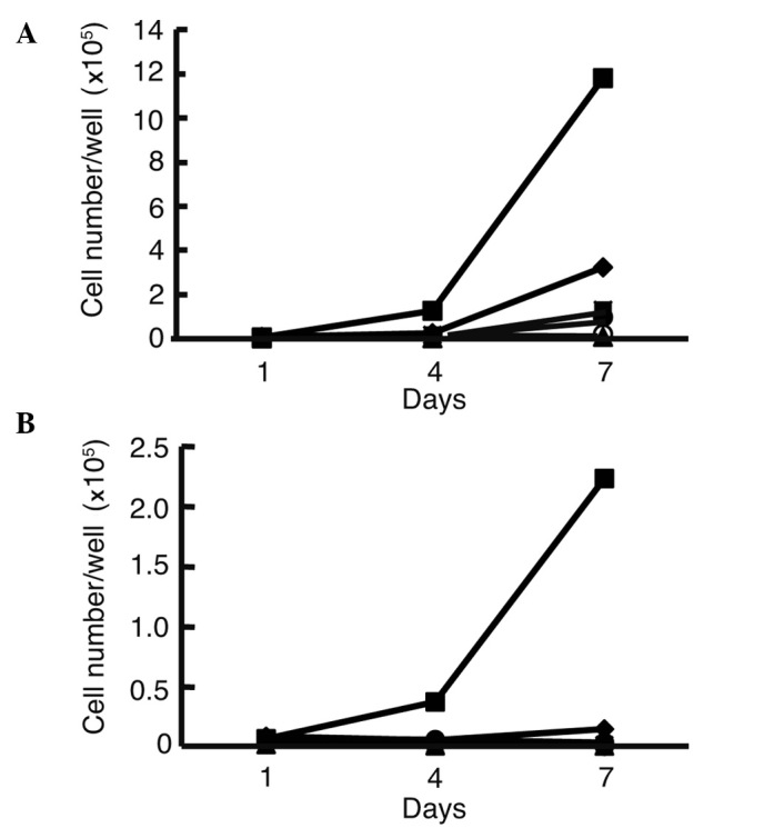
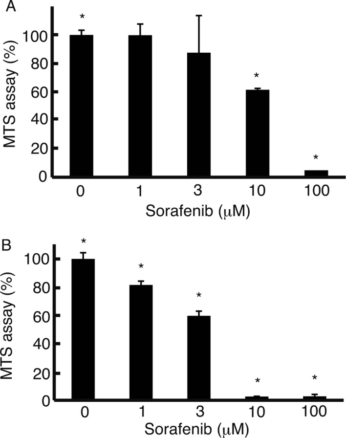
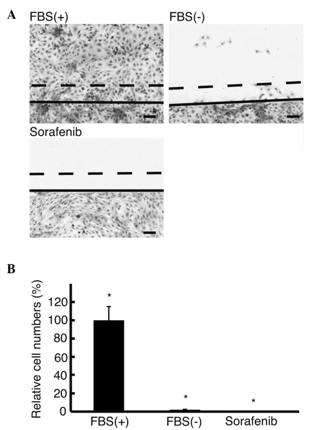
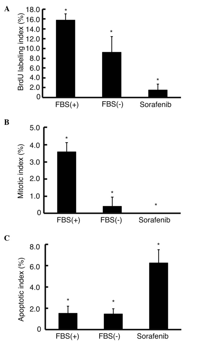
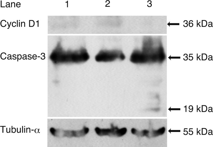
To suppress the invasion of hepatocellular carcinoma (HCC) cells into surrounding connective tissues during metastasis, we investigated the usefulness of sorafenib. In order to search for model cell lines, cell numbers were counted to reveal cell lines with the potential to proliferate in serum-free media. Cell proliferation and cell motility were analyzed with the MTS and wound assay, respectively. 5-Bromo-2'-deoxyuridine (BrdU) labeling and mitotic and apoptotic indices were analyzed to assess the cell cycle and apoptosis. The expression levels of cyclin D1 and the cleavage of caspase-3 were analyzed by Western blotting. HLF cells exhibited growth in the serum-free medium, while the other cell lines examined did not. Sorafenib suppressed the cell proliferation and motility of the HLF cells in the serum-free media. Both indices of BrdU and mitotic potential decreased and the apoptotic index was increased in the serum-free media with sorafenib, suggesting that the cell cycle was suppressed and apoptosis was induced. The expression levels of cyclin D1 decreased and the cleavage of caspase-3 was noted in the serum-free media with sorafenib. Sorafenib may be suitable for molecular therapy to suppress the metastasis of HCC.


Figures

Figure 1.
Figure 1.
Growth curve of hepatocellular carcinoma and hepatoblastoma cell lines. Cell numbers were counted 1, 4 and 7 days after the plating of 104 cells onto each well of 6-well plates in media with 10% FBS (A) or without FBS (B). HLE cells grew in medium with FBS, while HLF cells grew most rapidly in media without FBS. HLE (♦), HLF (▪), PLC/PRF/5 (▴), Huh-7 (x), Hep3B (○), Huh-6 (•) and HepG2 (⋄).
Figure 2.
Figure 2.
MTS assay. The MTS assay was performed to analyze the proliferation of HLF cells cultured in media with 10% FBS (A) and the proliferation of HLF cells in media without FBS (B) with sorafenib. Cell proliferation was suppressed as the concentration of sorafenib increased. In the media without FBS, cell proliferation was suppressed to a greater exent than the cell proliferation in the media with FBS, at the same concentrations of sorafenib. Error bar, standard deviation, *P<0.05, n=3.
Figure 3.
Figure 3.
Wound assay. (A) The wound assay was performed to analyze cell motility using H&E-stained culture slides. (B) The number of HLF cells migrating >150 μm per 100 μm of cut surface was counted. No cells migrated >150 μm in the media without FBS with sorafenib (Sorafenib). Solid line in A, edge of scratch; dotted line in A, 150 μm from the solid line. Original magnification, x4; scale bar, 100 μm. FBS(+), media with 10% FBS; FBS(−), media without FBS; Sorafenib, media without FBS with 10 μM sorafenib.
Figure 4.
Figure 4.
BrdU labeling, mitotic and apoptotic indices. (A) BrdU (10 μM) was added to the culture medium, and the number of positive cells was counted to analyze DNA synthesis immunohistochemically. (B) Mitotic cell number was counted using H&E-stained culture slides. (C) Apoptotic cell number was counted using culture slides with TUNEL assay. Sorafenib significantly decreased the numbers of both BrdU-positive and mitotic cells, while significantly increasing those of apoptotic cells in media without FBS (see text for the calculation of percentages). Error bar, standard deviation. *P<0.05, n=3. FBS(+), media with 10% FBS; FBS(−), media without FBS; Sorafenib, media without FBS with 10 μM sorafenib.
Figure 5.
Figure 5.
Western blot analysis. Western blot analysis was performed to analyze the molecular mechanism of the suppression of DNA synthesis and stimulation of apoptosis. The expression of cyclin D1 decreased (lane 2), while 19-kDa caspase-3, a cleaved form, was noted (lane 3) in cells in media with sorafenib. Lane 1: cells in media with 10% FBS; lane 2: cells in media without FBS; lane 3: cell in media without FBS with 10 μM sorafenib.

References

    1. Kondo Y, Kondo F, Wada K, Okabayashi A. Pathologic features of small hepatocellular carcinoma. Acta Pathol Jpn. 1986;36:1149–1161. - PubMed
    1. Tomizawa M, Kondo F, Kondo Y. Growth patterns and interstitial invasion of small hepatocellular carcinoma. Pathol Int. 1995;45:352–358. - PubMed
    1. Gupta GP, Massague J. Cancer metastasis: building a framework. Cell. 2006;127:679–695. - PubMed
    1. Takasu M, Tada Y, Wang JO, Tagawa M, Takenaga K. Resistance to apoptosis induced by microenvironmental stresses is correlated with metastatic potential in Lewis lung carcinoma. Clin Exp Metastasis. 1999;17:409–416. - PubMed
    1. Boraldi F, Annovi G, Paolinelli-Devincenzi C, Tiozzo R, Quaglino D. The effect of serum withdrawal on the protein profile of quiescent human dermal fibroblasts in primary cell culture. Proteomics. 2007;8:66–82. - PubMed