Review
. 2012:66:177-96.
doi: 10.1146/annurev-micro-092611-150203.

A virological view of innate immune recognition

Affiliations
Review

A virological view of innate immune recognition

Akiko Iwasaki. Annu Rev Microbiol. 2012.

Abstract

The innate immune system uses multiple strategies to detect viral infections. Because all viruses rely on host cells for their synthesis and propagation, the molecular features used to detect viral infections must be unique to viruses and absent from host cells. Research in the past decade has advanced our understanding of various cell-intrinsic and cell-extrinsic modes of virus recognition. This review examines the innate recognition from the point of view of virus invasion and replication strategies, and places innate sensors in the context of detecting viral genome, replication intermediate, transcriptional by-product, and other viral invasion strategies. On the basis of other unique features common to viral infections, undiscovered areas of virus detection are discussed.


Figures
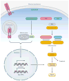
Figure 1
Figure 1
Pathways engaged following activation of innate viral sensors. TLRs reside either on the cell surface or in the endosomes; the latter requires cleavage for signaling. RLRs are present in the cytosol. Upon engagement of TLRs and RLRs by viruses, the receptor transmits signals that lead to the transcriptional activation of hundreds of genes including cytokines and type I IFNs. NLR and ALR proteins are localized in the cytosol. Certain virus infection leads to the activation of these receptors to form inflammasome, a large multimeric complex consisting of a subset of NLR/ALR, ASC, and pro-caspase-1. Caspase-1 becomes activated and cleaves its substrates including pro-IL-1β and pro-IL-18 for extracellular release. Cross talks between these pathways and exceptions are discussed throughout this review. Abbreviations: TLR, Toll-like receptor; RLR, RIG-I-like receptor; ALR, AIM2-like receptor; ASC, apoptosis-associated speck-like protein containing a caspase recruitment domain; MAVS, mitochondria antiviral signaling protein; NLR, Nod-like receptor; IL, interleukin; IFN, interferon.
Figure 2
Figure 2
Innate sensors and Baltimore classification of viruses. All viruses fall into one of seven groups depending on a combination of their genomes (DNA, RNA), strandedness (single or double), sense (sense or antisense), and mode of replication. This classification enables innate sensors to be placed into functional categories. TLRs, RLRs, and other sensors that recognize respective groups of viruses are indicated. Superscript a denotes sensors that have been identified by genetic knockdown studies, and superscript b denotes sensors associated with virus-induced diseases in humans. Abbreviations: TLR, Toll-like receptor; RLR, RIG-I-like receptor; mRNA, messenger RNA; Pol, polymerase; MDA5, melanoma differentiation-associated gene 5; RIG-I, retinoic-acid-inducible gene I; DAI, DNA-dependent activator of interferon-regulatory factors; IFI, interferon-inducible protein; DHX, DEAH box protein; LRRFIP, leucine-rich repeat flightless-interacting protein.
Figure 3
Figure 3
Known and putative viral PAMPs. Innate sensors can detect viral genomes in the endosomes ( purple boxes) or in the cytosol inside infected cells ( yellow boxes). Green letters denote cytosolic sensors, purple letters denote endosomal sensors, and blue letters denote antiviral effector ISGs. Host counterparts, where appropriate, are depicted at the bottom. Viral signatures predicted to serve as PAMPs are indicated by the pink boxes. These include sfRNA (Group IV), cap 0 structure of Sindbis virus mRNA, ssDNA in the nucleus (Group II), circular DNA (Group I, II), ssDNA and dsDNA in the cytosol (Group VI), and leader RNA (Group V). Abbreviations: PAMP, pathogen-associated molecular pattern; ISG, interferon-stimulated gene; sfRNA, subgenomic Flavivirus RNA; ssDNA, single-stranded DNA; dsDNA, double-stranded DNA; PKR, double-stranded RNA-activated protein kinase; OAS, 2′,5′-oligoadenylate synthase; RIG-I, retinoic-acid-inducible gene I; IFIT, interferon-induced tetratricopeptide repeat protein; MDA5, melanoma differentiation-associated protein 5; TREX1, three prime repair exonuclease 1; TLR, Toll-like receptor; DI, defective interfering; EBV, Epstein-Barr virus; EBER, EBV-encoded RNA; DHX, DEAH box protein; ppp, triphosphate; pA, poly(A) tail; Vpg, viral protein genome-linked; mRNA, messenger RNA; DAI, DNA-dependent activator of interferon-regulatory factors; AIM2, absent in melanoma 2; KSHV, Kaposi’s sarcoma-associated herpesvirus; IRES, internal ribosomal entry site; tRNA, transfer RNA.
Figure 4
Figure 4
Pol III viral transcripts activate RIG-I but inhibit PKR. Pol III transcripts generated during adenoviral infection (VA RNA) and EBV infection (EBER) are small, noncoding RNAs that bind to and block PKR activation. In the absence of viral infection, neither PKR nor RIG-I is activated. In cells infected with a virus (excluding adenovirus and EBV), dsRNA structure generated in the cytosol triggers the activation of PKR and 5′-ppp RNA triggers RIG-I activation, resulting in an antiviral state. In cells infected with adenovirus or EBV, noncoding RNA Pol III transcripts bind to RIG-I and stimulate IFN synthesis. However, Pol III transcripts bind to PKR and block its activity by disabling binding of stimulatory dsRNA. Abbreviations: Pol, polymerase; VA RNA, viral associated RNA; EBV, Epstein-Barr virus; EBER, EBV-encoded RNA; RIG-I, retinoic-acid-inducible gene 1; PKR, double-stranded RNA-activated protein kinase; dsRNA, double-stranded RNA; IFN, interferon.

Similar articles

Cited by

References

    1. Ablasser A, Bauernfeind F, Hartmann G, Latz E, Fitzgerald KA, Hornung V. RIG-I-dependent sensing of poly(dA:dT) through the induction of an RNA polymerase III-transcribed RNA intermediate. Nat Immunol. 2009;10:1065–72. Shows that Pol III transcripts from dsDNA viruses are recognized by RIG-I (see also Reference 18) - PMC - PubMed
    1. Akira S, Uematsu S, Takeuchi O. Pathogen recognition and innate immunity. Cell. 2006;124:783–801. - PubMed
    1. Alexopoulou L, Holt AC, Medzhitov R, Flavell RA. Recognition of double-stranded RNA and activation of NF-κB by Toll-like receptor 3. Nature. 2001;413:732–38. - PubMed
    1. Allen IC, Scull MA, Moore CB, Holl EK, McElvania-TeKippe E, et al. The NLRP3 inflammasome mediates in vivo innate immunity to influenza A virus through recognition of viral RNA. Immunity. 2009;30:556–65. - PMC - PubMed
    1. Andersson MG, Haasnoot PC, Xu N, Berenjian S, Berkhout B, Akusjarvi G. Suppression of RNA interference by adenovirus virus-associated RNA. J Virol. 2005;79:9556–65. - PMC - PubMed

Publication types

Substances

LinkOut - more resources