

Identification of amino acid residues important for anti-IFN activity of porcine reproductive and respiratory syndrome virus non-structural protein 1
- PMID: 22995188
- PMCID: PMC7111991
- DOI: 10.1016/j.virol.2012.08.034
Identification of amino acid residues important for anti-IFN activity of porcine reproductive and respiratory syndrome virus non-structural protein 1
Abstract
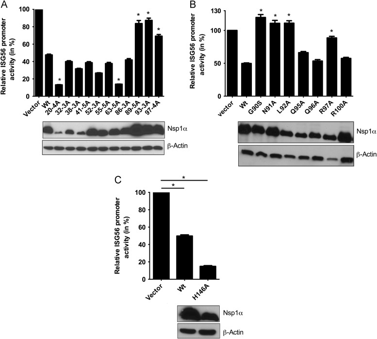
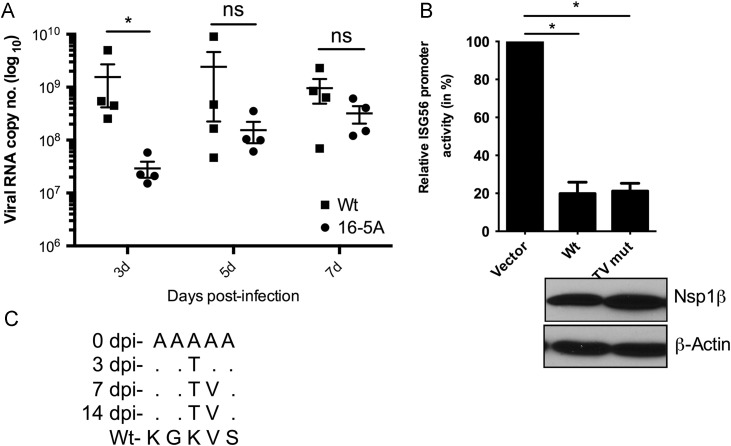
The non-structural protein 1 (nsp1) of porcine reproductive and respiratory syndrome virus is partly responsible for inhibition of type I interferon (IFN) response by the infected host. By performing alanine-scanning mutagenesis, we have identified amino acid residues in nsp1α and nsp1β (the proteolytic products of nsp1) that when substituted with alanine(s) exhibited significant relief of IFN-suppression. A mutant virus (16-5A, in which residues 16-20 of nsp1β were substituted with alanines) encoding mutant nsp1β recovered from infectious cDNA clone was shown to be attenuated for growth in vitro and induced significantly higher amount of type I IFN transcripts in infected macrophages. In infected pigs, the 16-5A virus exhibited reduced growth at early times after infection but quickly regained wild type growth properties as a result of substitutions within the mutated sequences. The results indicate a strong selection pressure towards maintaining the IFN-inhibitory property of the virus for successful propagation in pigs.
Copyright © 2012 Elsevier Inc. All rights reserved.
Figures
Similar articles
-
Mutations in a Highly Conserved Motif of nsp1β Protein Attenuate the Innate Immune Suppression Function of Porcine Reproductive and Respiratory Syndrome Virus.J Virol. 2016 Jan 20;90(7):3584-99. doi: 10.1128/JVI.03069-15. J Virol. 2016. PMID: 26792733 Free PMC article.
-
Amino acid residues in the non-structural protein 1 of porcine reproductive and respiratory syndrome virus involved in down-regulation of TNF-α expression in vitro and attenuation in vivo.Virology. 2012 Oct 25;432(2):241-9. doi: 10.1016/j.virol.2012.05.014. Epub 2012 Jun 13. Virology. 2012. PMID: 22699004
-
Type I interferon suppression-negative and host mRNA nuclear retention-negative mutation in nsp1β confers attenuation of porcine reproductive and respiratory syndrome virus in pigs.Virology. 2018 Apr;517:177-187. doi: 10.1016/j.virol.2018.01.016. Virology. 2018. PMID: 29402432
-
Interplay between interferon-mediated innate immunity and porcine reproductive and respiratory syndrome virus.Viruses. 2012 Apr;4(4):424-46. doi: 10.3390/v4040424. Epub 2012 Apr 2. Viruses. 2012. PMID: 22590680 Free PMC article. Review.
-
In vivo growth of porcine reproductive and respiratory syndrome virus engineered nsp2 deletion mutants.Virus Res. 2010 Dec;154(1-2):77-85. doi: 10.1016/j.virusres.2010.07.024. Epub 2010 Jul 29. Virus Res. 2010. PMID: 20673840 Free PMC article. Review.
Cited by
-
Infection dynamics, transmission, and evolution after an outbreak of porcine reproductive and respiratory syndrome virus.Front Microbiol. 2023 Feb 9;14:1109881. doi: 10.3389/fmicb.2023.1109881. eCollection 2023. Front Microbiol. 2023. PMID: 36846785 Free PMC article.
-
Equine arteritis virus does not induce interferon production in equine endothelial cells: identification of nonstructural protein 1 as a main interferon antagonist.Biomed Res Int. 2014;2014:420658. doi: 10.1155/2014/420658. Epub 2014 May 25. Biomed Res Int. 2014. PMID: 24967365 Free PMC article.
-
Functional analyses of the three simian hemorrhagic fever virus nonstructural protein 1 papain-like proteases.J Virol. 2014 Aug;88(16):9129-40. doi: 10.1128/JVI.01020-14. Epub 2014 Jun 4. J Virol. 2014. PMID: 24899184 Free PMC article.
-
Porcine Reproductive and Respiratory Syndrome Virus Reverse Genetics and the Major Applications.Viruses. 2020 Oct 31;12(11):1245. doi: 10.3390/v12111245. Viruses. 2020. PMID: 33142752 Free PMC article. Review.
-
Characterization of a serologic marker candidate for development of a live-attenuated DIVA vaccine against porcine reproductive and respiratory syndrome virus.Vaccine. 2013 Sep 13;31(40):4330-7. doi: 10.1016/j.vaccine.2013.07.020. Epub 2013 Jul 23. Vaccine. 2013. PMID: 23892102 Free PMC article.
References
-
- Albina E., Carrat C., Charley B. Interferon-alpha response to swine arterivirus (PoAV), the porcine reproductive and respiratory syndrome virus. J. Interferon Cytokine Res. 1998;18(7):485–490. - PubMed
Publication types
MeSH terms
Substances
LinkOut - more resources
Full Text Sources
Other Literature Sources
Research Materials
