. 2013 Mar;44(3):374-87.
doi: 10.1016/j.humpath.2012.03.031. Epub 2012 Sep 17.

Expression of the vitamin D-activating enzyme 1α-hydroxylase (CYP27B1) decreases during melanoma progression

Affiliations

Expression of the vitamin D-activating enzyme 1α-hydroxylase (CYP27B1) decreases during melanoma progression

Anna A Brożyna et al. Hum Pathol. 2013 Mar.

Abstract

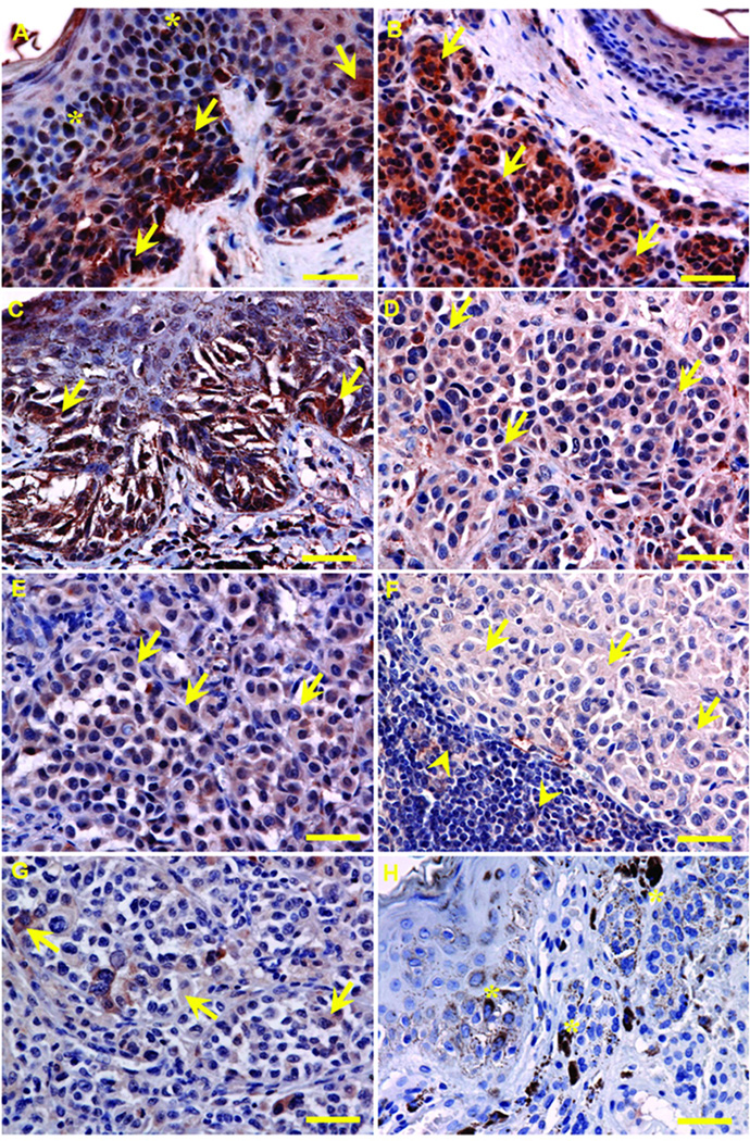
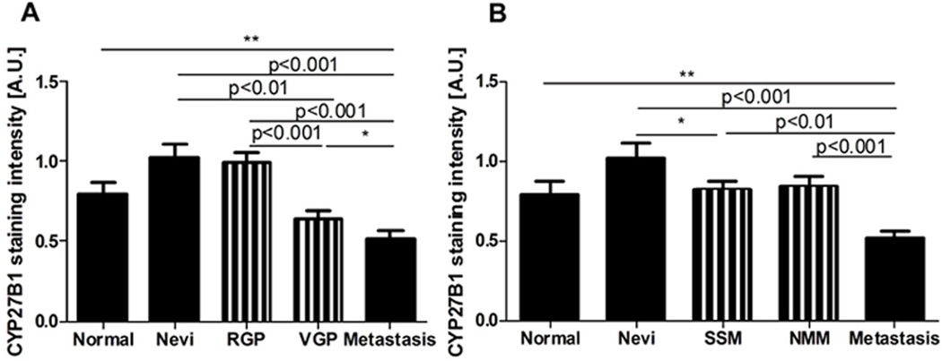
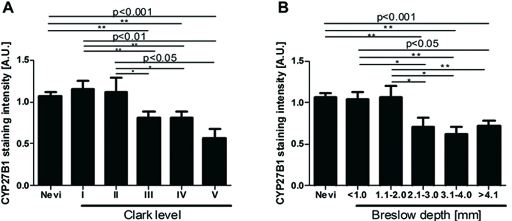
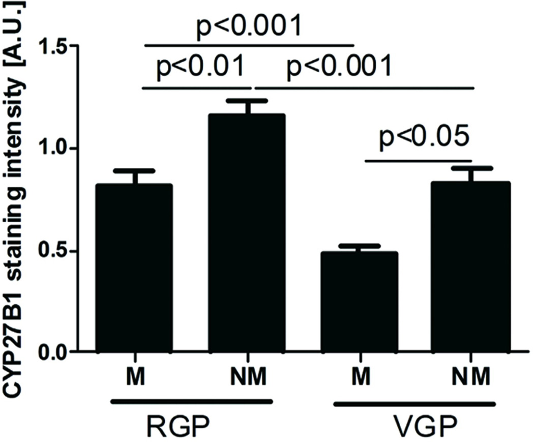
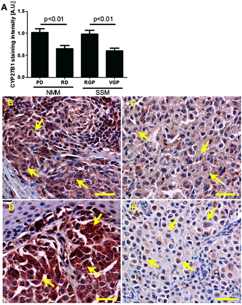
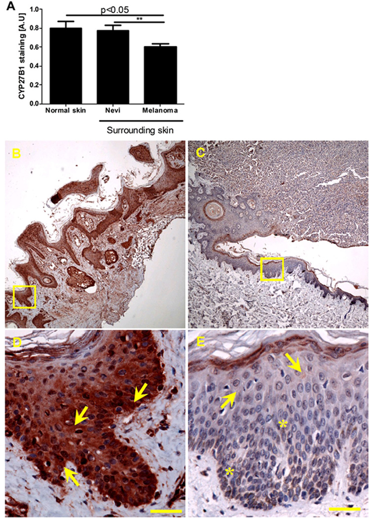
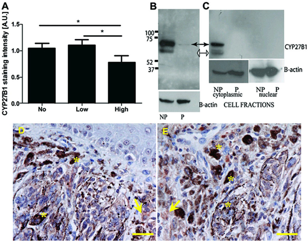
1α-Hydroxylase (CYP27B1), the enzyme responsible for the synthesis of the biologically active form of vitamin D (1,25(OH)(2)D(3)), is expressed in the skin. To assess the correlation between progression of melanocytic tumors and CYP27B1, we analyzed its expression in 29 benign nevi, 75 primary cutaneous melanomas, 40 metastases, and 4 re-excision and 6 normal skin biopsies. Immunoreactivity for CYP27B1 was significantly lower in the vertical growth phase and metastatic melanomas (0.6 and 0.5 arbitrary units, respectively) in comparison with nevi and radial growth phase tumors (1.2 and 1.1 arbitrary units, respectively); and expression was reduced in more advanced lesions (Clark levels III-V, Breslow thickness ≥2.1 mm; 0.8 and 0.7 arbitrary units, respectively). There was an inverse correlation between CYP27B1 and Ki-67 expression. Furthermore, CYP27B1 expression was reduced in primary melanomas that created metastases in comparison with non-metastasizing melanomas. Reduced CYP27B1 expression in radial growth phase was related to shorter overall survival (810 versus 982 versus 1151 days in melanomas with absent, low, and high CYP27B1 immunoreactivity), and low CYP27B1 expression in radial growth phase and vertical growth phase was related to shorter disease-free survival (114 versus 339 versus 737 days and 129 versus 307 versus 737 days, respectively, in melanomas with absent, low, and high CYP27B1). Also, CYP27B1 expression was inversely related to melanin in melanoma cells in vivo and melanoma cells cultured in vitro. Thus, reduction of CYP27B1 correlates with melanoma phenotype and behavior, and its lack affects the survival of melanoma patients, indicating a role in the pathogenesis and progression of this cancer.


Figures

Figure 1
Figure 1
Representative CYP27B1 immunostaining in benign junctional (A) and dermal (B) melanocytic nevi, melanoma in situ (C), invasive superficial spreading (SSM) (D) and nodular (NMM) (E) melanomas, lymph node metastases (F), and recurrent melanomas (G). Nevus sections immunoprocessed without primary antibodies were the negative control (H). Arrows indicate CYP27B1 immunostaining; arrowheads mark lymphocytes, asterisks show melanin. Scale bar: 50 µm.
Figure 2
Figure 2
Changes in expression of CYP27B1 in normal skin, melanocytic nevi, and metastases in comparison with melanomas classified according growth phase (A) or histologic type (B). Radial growth phase (RGP), vertical growth phase (VGP), superficial spreading (SSM), and nodular (NMM) melanomas. Statistically significant differences are denoted with P values after ANOVA and with asterisks as *P < .05 or **P < .01 with t test.
Figure 3
Figure 3
CYP27B1 expression in cytoplasm of nevi and melanomas stratified according to Clark level (A) and Breslow thickness (B). Statistically significant differences are denoted with P values after ANOVA and with asterisks as *P < .05, **P < .01, and ***P < .001 with t test.
Figure 4
Figure 4
CYP27B1 immunostaining in metastasizing (M) and non-metastasizing (NM) melanomas. Statistically significant differences are denoted with P values after ANOVA.
Figure 5
Figure 5
Expression of CYP27B1 decreases during melanoma progression. Comparison of CYP27B1 immunostaining in RGP and VGP of SSM and in papillary (PD) and reticular (RD) dermis compartments of NMM (A). Representative CYP27B1 immunostaining of RGP (B) and VGP (C) of SSM and in PD (D) and RD (E) compartments of NMM. Statistically significant differences are denoted with P values after ANOVA. Arrows indicate CYP27B1 expression. Scale bar: 50 µm.
Figure 6
Figure 6
Comparison of CYP27B1 immunostaining in normal skin and skin surrounding nevi and melanomas. Quantification of relative expression is in panel A, whereas representative immunostains for CYP27B1 in normal skin and skin surrounding melanomas are in panels B and C, respectively. D–E, High magnification views of areas indicated in squares in normal skin (D) and skin surrounding melanoma (E). Statistically significant differences are denoted with P values after ANOVA and with asterisks as *P < .05 with t test. Arrows indicate CYP27B1 staining. Scale bar: 50 µm.
Figure 7
Figure 7
Proliferative activity in melanomas in RGP (A) and VGP (B) shows inverse correlation with expression of CYP27B1. Only one case of melanoma in VGP had high CYP27B1 immunostaining. Statistically significant differences are denoted by asterisks as **P < .01 with t test.
Figure 8
Figure 8
Correlation of overall survival time (A) and diseases-free survival time (B and C) with relative expression of CYP27B1. A and B represents RGP, and C represents VGP.
Figure 9
Figure 9
Expression of CYP27B1 mRNA in melanoma lines in comparison with normal human keratinocytes (HEKn) (A) or melanocytes (HEMn) (B) measured by real-time RT PCR. Data are presented as mean ± SD (n = 3). Statistically significant differences are denoted with asterisks as *P < .05, **P < .01, and ***P < .001 with t test.
Figure 10
Figure 10
CYP27B1 expression decreases in highly melanized melanoma cells. CYP27B1 expression in melanoma samples measured by immunocytochemistry. Statistically significant differences are denoted with P values after ANOVA and with asterisks as *P < .05 with t test (A). Induction of melanin synthesis in cultured SKMel-188 melanoma cells reduced the CYP27B1 as measured by Western blotting (B and C). Whole extracts from nonpigmented (NP) and pigmented (P) cells were subjected to immunoblotting with anti-CYP27B1 and anti-β-actin (internal control)(B). Isolated nuclear and cytoplasmic extracts were subjected to immunoblotting with anti-CYP27B1 and anti-β-actin (internal control)(C). Representative CYP27B1 immunostaining of RGP (D) and VGP (E) pigmented melanoma. Arrows mark CYP27B1 staining; asterisks indicate melanin. Scale bar: 50 µm.

Similar articles

Cited by

References

    1. Wang TT, Tavera-Mendoza LE, Laperriere D, Libby E, MacLeod NB, Nagai Y, et al. Large-scale in silico and microarray-based identification of direct 1,25-dihydroxyvitamin D3 target genes. Mol Endocrinol. 2005;19:2685–2695. - PubMed
    1. Bikle DD. The vitamin D receptor: a tumor suppressor in skin. Discov Med. 2011;11:7–17. - PMC - PubMed
    1. Chen H, Hu B, Allegretto EA, Adams JS. The vitamin D response element-binding protein: a novel dominant-negative regulator of vitamin D-directed transactivation. J Biol Chem. 2000;275:35557–35564. - PubMed
    1. Colston K, Colston MJ, Feldman D. 1,25-Dihydroxyvitamin D3 and malignant melanoma: the presence of receptors and inhibition of cell growth in culture. Endocrinology. 1981;108:1083–1086. - PubMed
    1. Fleet JC. Molecular actions of vitamin D contributing to cancer prevention. Mol Aspects Med. 2008;29:388–396. - PMC - PubMed

Publication types

MeSH terms