

Immunohistochemistry with apoptotic-antiapoptotic proteins (p53, p21, bax, bcl-2), c-kit, telomerase, and metallothionein as a diagnostic aid in benign, borderline, and malignant serous and mucinous ovarian tumors
- PMID: 22995373
- PMCID: PMC3523067
- DOI: 10.1186/1746-1596-7-124
Immunohistochemistry with apoptotic-antiapoptotic proteins (p53, p21, bax, bcl-2), c-kit, telomerase, and metallothionein as a diagnostic aid in benign, borderline, and malignant serous and mucinous ovarian tumors
Abstract
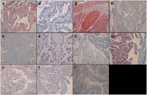
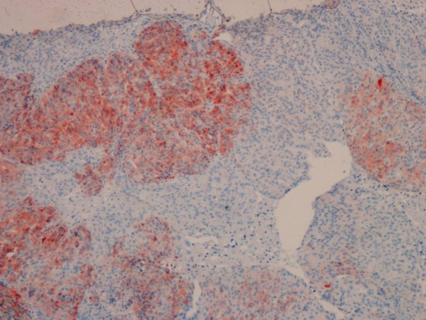
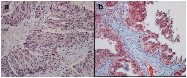
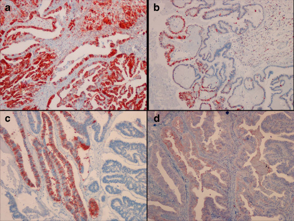
Background: In many tumors including ovarian cancer, cell proliferation and apoptosis are important in pathogenesis and there are many alterations in most of the genes related to the cell cycle. This study was designed to evaluate immunohistochemistry with apoptotic-antiapoptotic proteins (p53, p21, bax, and bcl-2), c-kit, telomerase, and metallothionein as a diagnostic aid in typing of benign, borderline, and malignant serous and mucinous ovarian tumors.
Methods: Total of 68 ovarian tumors, 25 benign [13 (19.1%) serous and 12 (17.6%) mucinous], 16 borderline [9 (13.2%) serous and 7(10.3%) mucinous], and 27 malignant ovarian tumors [24 (35.3%) serous and 3 (4.4%) mucinous tumors] were included in the study. Immunohistochemical expression of p53, p21, bax, bcl-2, telomerase, c-kit, and metallothionein were evaluated.
Results: When all 68 cases were evaluated as benign, borderline, and malignant ovarian tumors without considering histopathological subtypes, the p53, p21, bax and metallothionein showed significantly higher staining scores in the borderline and malignant ones (p < 0.05). After evaluation of all 68 cases, the serous tumors showed significantly higher staining scores of p53, p21, c-kit, and metallothionein compared to the mucinous ones (p < 0.05). For differentiation of benign and borderline and malignant tumors combined, p53 was not used because all benign tumors has no staining, and p21, bax, and metallothionein was determined the significant predictors for borderline and malignant tumors combined (p < 0.05). For differentiation of borderline and malignant tumors, only p53 was determined the significant predictor for malignant tumors (p < 0.05).
Conclusions: In conclusion, p53, p21, bax, c-kit, and metallothionein may be helpful for the typing of ovarian tumors as benign, borderline and malignant or serous and mucinous. p53, p21, bax, c-kit, and metallothionein may have different roles in the pathogenesis of ovarian tumor types. p53 and metallothionein may be helpful in the typing of borderline and malignant ovarian tumors. The immunohistochemical staining with bcl-2 and telomerase may not provide meaningful contribution for the typing of ovarian tumors.
Virtual slide: The virtual slides for this article can be found here: http://www.diagnosticpathology.diagnomx.eu/vs/2013030833768498.
Figures
References
-
- Omura G, Blessing JA, Ehrlich CE, Miller A, Yordan E, Creasman WT, Homesley HD. A randomized trial of cyclophosphamide and doxorubicin with or without cisplatin in advanced ovarian carcinoma. A Gynecologic Oncology Group Study Cancer. 1986;57(9):1725–1730. - PubMed
-
- Coleman MP, Forman D, Bryant H, Butler J, Rachet B, Maringe C, Nur U, Tracey E, Coory M, Hatcher J, McGahan CE, Turner D, Marrett L, Gjerstorff ML, Johannesen TB, Adolfsson J, Lambe M, Lawrence G, Meechan D, Morris EJ, Middleton R, Steward J, Richards MA. ICBP Module 1 Working Group. Cancer survival in Australia, Canada, Denmark, Norway, Sweden, and the UK, 1995–2007 (the International Cancer Benchmarking Partnership): an analysis of population-based cancer registry data. Lancet. 2011;377(9760):127–38. doi: 10.1016/S0140-6736(10)62231-3. - DOI - PMC - PubMed
-
- Seidman JD, Russel P, Kurman RJ. In: In Blaustein’s Pathology of the Female Genital Tract. 5. Kurman RJ, editor. New York: Springer; 2002. Surface epithelial tumors of the ovary, (Chapter 18) pp. 791–904.
-
- Lee KR, Tavassoli FA, Prat J, Dietel M, Gersell DJ, Karseladze AI, Hauptmann S, Rutgers J. In: In Pathology and Genetics of Tumours of the Breast and Female Genital organs. Tavassoli FA, Devilee P, editor. Lyon: IARC Pres; 2003. WHO Histological Classification of Tumours of the ovary (Chapter 2) pp. 113–161.
Publication types
MeSH terms
Substances
LinkOut - more resources
Full Text Sources
Medical
Research Materials
Miscellaneous
