. 2012 Nov;32(11):2598-608.
doi: 10.1161/ATVBAHA.112.300310. Epub 2012 Sep 20.

Endogenous IRAK-M attenuates postinfarction remodeling through effects on macrophages and fibroblasts

Affiliations

Endogenous IRAK-M attenuates postinfarction remodeling through effects on macrophages and fibroblasts

Wei Chen et al. Arterioscler Thromb Vasc Biol. 2012 Nov.

Abstract

Objective: Effective postinfarction repair requires timely suppression of innate immune signals to prevent the catastrophic consequences of uncontrolled inflammation on cardiac geometry and function. In macrophages, interleukin-1 receptor-associated kinase (IRAK)-M acts as a functional decoy preventing uncontrolled toll-like receptor /interleukin-1-mediated responses. Our study investigates the role of IRAK-M as a negative regulator of the postinfarction inflammatory response and as a modulator of cardiac remodeling.

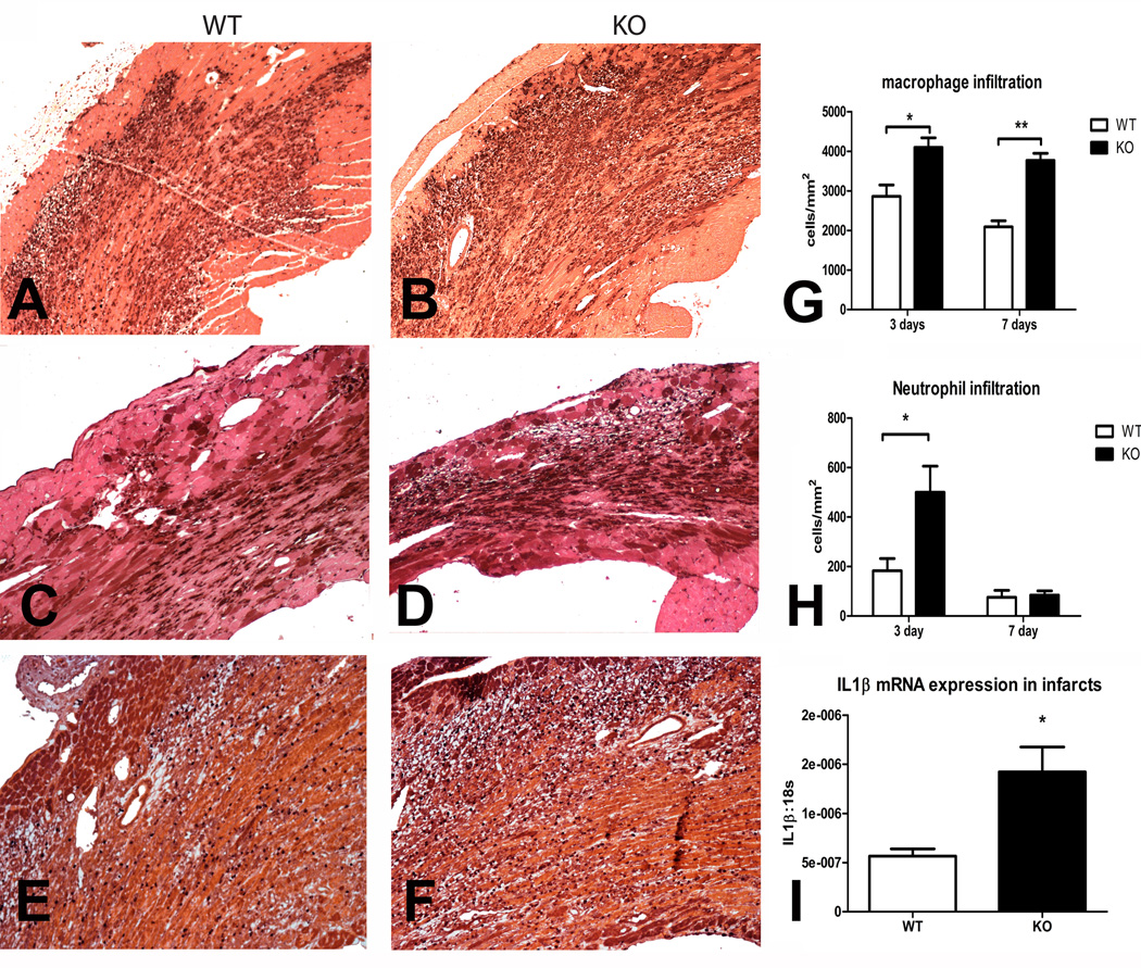
Methods and results: In wild-type mouse infarcts IRAK-M was upregulated in infiltrating macrophages and fibroblasts exhibiting a biphasic response. When compared with wild-type animals, infarcted IRAK-M(-/-) mice had enhanced adverse remodeling and worse systolic dysfunction; however, acute infarct size was comparable between groups. Adverse remodeling in IRAK-M(-/-) animals was associated with enhanced myocardial inflammation and protease activation. The protective actions of IRAK-M involved phenotypic modulation of macrophages and fibroblasts. IRAK-M(-/-) infarcts showed increased infiltration with proinflammatory CD11b+/Ly6C(hi) monocytes; leukocytes harvested from IRAK-M-null infarcts exhibited accentuated cytokine expression. In vitro, IRAK-M expression was upregulated in cytokine-stimulated murine cardiac fibroblasts and suppressed their matrix-degrading properties without affecting their inflammatory activity.

Conclusions: Endogenous IRAK-M attenuates adverse postinfarction remodeling suppressing leukocyte inflammatory activity, while inhibiting fibroblast-mediated matrix degradation.


Figures

Figure 1
Figure 1
IRAK-M upregulation in the infarcted mouse heart. A. IRAK-M mRNA expression in the infarcted myocardium showed a biphasic response: significant upregulation was noted after 6h of reperfusion followed by a second peak after 7 days of reperfusion (**p<0.01 versus sham). B–C. Dual immunofluorescence combining IRAK-M staining (red) and Mac-2 immunofluorescence (B, green) to identify macrophages, or α-SMA staining to label myofibroblasts and smooth muscle cells (C, green). Dual immunofluorescent staining localized IRAK-M (red) in Mac2+ macrophages (B- arrows) and spindle-shaped α-smooth muscle actin+ myofibroblasts (C- arrows) infiltrating the infarcted myocardium (1h ischemia/7 days reperfusion). Counterstained with DAPI (blue). D-G. Fibroblasts (D) and macrophages (E) isolated from infarcted hearts 3 days after reperfusion expressed IRAK-M (green, arrows). IRAK-M immunofluorescence in fibroblasts (F) and macrophages (G) isolated from IRAK-M null infarcts served as a negative control. Cells were counterstained with DAPI (blue). H–J. IRAK-M loss did not affect the size of the infarct after 1h ischemia and 24h of reperfusion. TTC/Evans Blue staining was used to measure the area at risk (AAR) and the infarcted area (INF) in the ischemic and reperfused heart. WT and IRAK-M null animals had comparable AAR (H), infarcted area (I) and INF:AAR ratio (J).
Figure 2
Figure 2
Echocardiography (A–G) and quantitative morphometry (H–L) demonstrate that IRAK-M absence results in accentuated dilative remodeling and worse systolic dysfunction following reperfused myocardial infarction. A. Representative images of long axis B-mode and short axis M-mode echocardiography in WT and IRAK-M KO mice at baseline and after 7–28 days of reperfusion. B–G: IRAK-M null mice had worse systolic dysfunction and accentuated remodeling of the infarcted heart when compared with WT animals. Quantitative analysis of echocardiographic parameters demonstrated that IRAK-M null hearts had increased chamber dilation (indicated by higher LVEDD, LVESD, LVEDV and LVESV; B, C, E, F), worse systolic dysfunction (indicated by lower FS, D) and accentuated hypertrophy (evidenced by higher LV mass, G) after 7–28 days of reperfusion (**p<0.01, *p<0.05 vs. corresponding WT). H. Quantitative morphometric analysis demonstrates that IRAK-M disruption is associated with increased post-infarction remodeling. Hearts were perfusion-fixed and systematic morphometric analysis of the geometry of the left ventricle was performed through assessment of sections cut at 250µm intervals from base to apex. Representative images show cross-sections of WT and IRAK-M null sham and infarcted hearts (after 7 and 28 days of reperfusion). I-L: Quantitative analysis demonstrated that, although scar size was comparable between IRAK-M null and WT animals after 7–28 days of reperfusion (B), IRAK-M −/− mice had increased LVEDD and LVEDV and higher LV mass when compared with WT animals (**p<0.01, *p<0.05 vs. corresponding WT mice).
Figure 3
Figure 3
IRAK-M null mice exhibit an accentuated inflammatory response following myocardial infarction. A–D. Representative images show identification of macrophages in the infarcted myocardium using Mac-2 immunohistochemistry after 72h (A–B) and 7 days of reperfusion (C–D) E–F: Representative images show immunohistochemical staining with an anti-neutrophil antibody after 72h of reperfusion. G: Quantitative analysis showed that macrophage density was significantly higher in IRAK-M null infarcts (**p<0.01, *p<0.05 vs. corresponding WT). H: Neutrophil density was markedly higher in IRAK-M null infarcts after 72h of reperfusion; however, resolution of the neutrophil infiltrate occurred in a timely manner in both groups. I: IL-1β mRNA levels were markedly higher in IRAK-M null infarcts suggesting accentuation of the inflammatory response. All sections were counterstained with eosin.
Figure 4
Figure 4
Adverse remodeling in infarcted IRAK-M−/− hearts is associated with increased MMP mRNA expression and enhanced protease activity. A–F: qPCR demonstrated increased expression of MMPs (A, MMP-2; B, MMP-3; C, MMP-8; D, MMP-9) and TIMPs (E, TIMP-1; F, TIMP-2) in IRAK-M null infarcts after 24h of reperfusion. G: Zymography was used to quantitatively assess MMP activity in the infarcted myocardium after 72h of reperfusion. H: Quantitative analysis demonstrated that levels of active MMP-2 were markedly higher in IRAK-M null infarcts (**p<0.01 vs. WT). A trend towards increased MMP-9 activity was also noted (p=0.19). I-J: Picrosirius red staining was used to assess collagen deposition in the scar (1hI /7 days R). Increased MMP activity in IRAK-M −/− mice was associated with less collagen deposition. (**p<0.01, *p<0.05 vs. corresponding WT).
Figure 5
Figure 5
A. IRAK-M regulation in stimulated cardiac fibroblasts. IL-1β, the TLR agonist LPS, TNF-α, and PDGF-BB upregulated IRAK-M mRNA expression in cardiac fibroblasts (**p<0.01, *p<0.05 vs. control). mRNA isolated from IRAK-M −/− fibroblasts was used as a negative control. B–C. IRAK-M loss enhances cytokine-stimulated MMP expression, but does not affect inflammatory cytokine synthesis in isolated cardiac fibroblasts. B. IRAK-M −/− fibroblasts exhibited increased MMP-2 and MMP-8 expression upon stimulation with IL-1β, when compared with WT fibroblasts. In contrast, MMP-3 and TIMP-1 expression was comparable between groups. C. IL-1β-mediated upregulation of TNF-α and MCP-1 was comparable between WT and IRAK-M null fibroblasts. IRAK-M null fibroblasts had reduced IL-6 upregulation upon IL-1β stimulation.
Figure 6
Figure 6
IRAK-M loss is associated with infiltration of the infarcted myocardium with pro-inflammatory monocytes. Flow cytometric analysis of cell suspensions harvested from the infarcted heart after 72h of reperfusion demonstrated marked increases in the number of infiltrating mononuclear cells and IL1β-positive cells. Cell suspensions from infarcted hearts of C57BL/6 and IRAK-M null mice were stained with LIVE/DEAD® Fixable Dead Cell Stain, -CD45, -CD11b, -Ly6C, -F4/80 and -IL1β mAbs. A–B: Representative dot plots show significantly higher number of live CD45+ hematopoietic cells (A), CD11b+/F4/80- monocytes (gate 1) and CD11b+F4/80+ macrophages/dendritic cells (gate 2) (B) in IRAK-M−/− infarcts after 72hr of reperfusion. C: Representative dot plots show the percentage of IL1β positive cells in Ly6Chi and Ly6Clo monocytes. D: IL1β expression in Ly6Clo and Ly6Chi subgroups of monocytic cells is shown in representative histograms comparing findings from WT (blue) and IRAK-M null infarcts (red). E–L: Quantitative analysis of flow cytometric findings showed that IRAK-M −/− infarcts had markedly higher absolute number of CD45+ hematopoietic cells (E), CD45+/CD11b+ monocytic cells (F), Ly6Chi/CD45+/CD11b+ pro-inflammatory monocytes (G) and F4/80+ macrophages (H) per mg of weight of the infarcted heart. Monocytes and macrophage subpopulations harvested from IRAK-M −/− infarcts had increased expression of the pro-inflammatory cytokine IL-1β reflecting accentuated inflammatory activity. The number of IL-1β+/CD45+ hematopoietic cells (I), IL-1β+/CD45+/CD11b+ monocytic cells (J), IL-1β+/CD11b+/Ly6Chi pro-inflammatory monocytes (K), and IL-1β+/F4/80+ macrophages (L) was markedly higher in IRAK-M null infarcts (**p<0.01, *p<0.05 vs. corresponding WT animals).

References

    1. Nathan C, Ding A. Nonresolving inflammation. Cell. 2010;140:871–882. - PubMed
    1. Maskrey BH, Megson IL, Whitfield PD, Rossi AG. Mechanisms of resolution of inflammation: a focus on cardiovascular disease. Arterioscler Thromb Vasc Biol. 2011;31:1001–1006. - PubMed
    1. Frangogiannis NG. Regulation of the inflammatory response in cardiac repair. Circ Res. 2012;110:159–173. - PMC - PubMed
    1. Pfeffer MA, Pfeffer JM. Ventricular enlargement and reduced survival after myocardial infarction. Circulation. 1987;75:IV93–IV97. - PubMed
    1. Holmes JW, Borg TK, Covell JW. Structure and mechanics of healing myocardial infarcts. Annu Rev Biomed Eng. 2005;7:223–253. - PubMed

Publication types

MeSH terms