. 2012 Nov 16:1485:95-107.
doi: 10.1016/j.brainres.2012.08.037. Epub 2012 Aug 24.

Multi-sensory integration in brainstem and auditory cortex

Affiliations

Multi-sensory integration in brainstem and auditory cortex

Gregory J Basura et al. Brain Res. .

Abstract

Tinnitus is the perception of sound in the absence of a physical sound stimulus. It is thought to arise from aberrant neural activity within central auditory pathways that may be influenced by multiple brain centers, including the somatosensory system. Auditory-somatosensory (bimodal) integration occurs in the dorsal cochlear nucleus (DCN), where electrical activation of somatosensory regions alters pyramidal cell spike timing and rates of sound stimuli. Moreover, in conditions of tinnitus, bimodal integration in DCN is enhanced, producing greater spontaneous and sound-driven neural activity, which are neural correlates of tinnitus. In primary auditory cortex (A1), a similar auditory-somatosensory integration has been described in the normal system (Lakatos et al., 2007), where sub-threshold multisensory modulation may be a direct reflection of subcortical multisensory responses (Tyll et al., 2011). The present work utilized simultaneous recordings from both DCN and A1 to directly compare bimodal integration across these separate brain stations of the intact auditory pathway. Four-shank, 32-channel electrodes were placed in DCN and A1 to simultaneously record tone-evoked unit activity in the presence and absence of spinal trigeminal nucleus (Sp5) electrical activation. Bimodal stimulation led to long-lasting facilitation or suppression of single and multi-unit responses to subsequent sound in both DCN and A1. Immediate (bimodal response) and long-lasting (bimodal plasticity) effects of Sp5-tone stimulation were facilitation or suppression of tone-evoked firing rates in DCN and A1 at all Sp5-tone pairing intervals (10, 20, and 40 ms), and greater suppression at 20 ms pairing-intervals for single unit responses. Understanding the complex relationships between DCN and A1 bimodal processing in the normal animal provides the basis for studying its disruption in hearing loss and tinnitus models. This article is part of a Special Issue entitled: Tinnitus Neuroscience.


Figures

Figure 1
Figure 1
A. Magnified histologic region of A1 demonstrating fluorogold labeling and tract tracing confirming Michigan Probe electrode insertion in A1 (left; A1, primary auditory cortex; ERF, entorhinal fissure). B. Schematic demonstrating electrode array placement in the dorsal cochlear nucleus (DCN) in a rostral-caudal dorsal-ventral plane from the surface of the DCN and stimulating current probe placement within the spinal trigeminal nucleus (Sp5) for somatosensory stimulation. C. Surgical preparation showing simultaneous placement of probes in DCN (left; ipsilateral to sound source), Sp5 (middle; ipsilateral to sound source) and A1 (right; contralateral to sound source).
Figure 2
Figure 2
Pairing protocol used to ascertain responses to immediate bimodal stimulation and long-lasting (5–10 minutes after pairing), persistent responses to sound following pairing. Responses to single level tones (Tone) were recorded during and after bimodal stimulation (pairing protocol). Recording blocks were randomly performed with varied pairing intervals between Sp5 always preceding tone (Pairing Intervals = 10, 20 or 40ms).
Figure 3
Figure 3
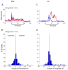
The immediate effect of bimodal stimulation on DCN(A) and A1(B). The far left hand column in both DCN and A1 demonstrates peri-stimulus time histograms (PSTH) showing the immediate effects of somatosensory-auditory stimulation (bimodal; red) as compared to sound alone (blue; middle column) at various pairing intervals (40, 20 and 10ms from top to bottom). Histograms in the far-right hand column of both groups reveal the corresponding quantified bimodal effect presented as a percentage of units with values right of zero (vertical line) being facilitated and those left of zero being suppressed at each pairing interval. Black bar under PSTHs indicates tone duration.
Figure 4
Figure 4
Bimodal stimulation results in long-lasting changes in DCN (A, B) and A1 (C, D). Upper panels (A and C) are peri-stimulus time histograms (PSTHs) demonstrating the long-lasting effects of somatosensory-auditory stimulation. Pre-bimodal responses are shown in blue; post-pairing responses to sound alone are shown in red. (Pairing interval = 10ms). Histograms for DCN (B) and A1 (D) demonstrate the long-term changes in firing rates following bimodal stimulation at a pairing interval of 10ms. Responses to the right of zero are facilitated, while those left of zero are suppressed. Black bar under PSTHs indicates tone duration.
Figure 5
Figure 5
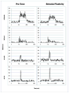
Peri-stimulus time histograms (PSTH) of responses from single and multi-unit analysis from DCN and A1 ((DCN-SU; A1-SU; DCN-MU; A1-MU). Long-term effects (Bimodal Plasticity; right column) of somatosensory-auditory stimulation are compared to sound alone (Pre-Tone; left column) for a 20ms pairing interval. Note the similarities in PSTH profiles between multi- and single-unit responses from both brain regions. Black bars under PSTHs indicate tone durations.
Figure 6
Figure 6
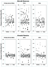
Mean effects of immediate (A; bimodal response) and long-lasting (B; bimodal plasticity) bimodal stimulation in DCN and A1. The percent change in firing rate after bimodal stimulation is plotted at each bimodal pairing interval. Values above zero represent facilitation; values below zero represent suppression. Single-unit responses are plotted for DCN (open circles) and A1 (filled-circles). Similar responses occur for single- and multi-units at all pairing-intervals in both brain stations with the exception of maximal suppression at 20ms in both bimodal response and plasticity.
Figure 7
Figure 7
Scatter plot demonstrating the immediate (Bimodal response; top) and long-lasting (Bimodal plasticity, bottom) effects of bimodal stimulation in A1 as a function of cortical depth at each pairing interval. The percent of units that are facilitated or suppressed are shown across cortical depths from shallow to deep. No significant trend is seen for facilitated or suppressed units across cortical depth. Single-units (filled triangles); Mulitunits (circles).
Figure 8
Figure 8
Anatomic diagram demonstrating the “already-processed” lemniscal auditory pathway and Sp5 innervation to the DCN. A secondary/”alternate” somatosensory pathway includes Sp5 projections to thalamus to somatosensory cortices and the auditory cortex belt region.

References

    1. Bauer CA, Turner JG, Caspary DM, Myers KS, Brozoski TJ. Tinnitus and inferior colliculus activity in chinchillas related to three distinct patterns of cochlear trauma. J Neurosci Res. 2008;15:2564–2578. - PMC - PubMed
    1. Brozoski T, Bauer C, Caspary D. Elevated fusiform cell activity in the dorsal cochlear nucleus of chinchillas with psychophysical evidence of tinnitus. PG - 2383–90. J Neurosci. 2002;22:2383–2390. - PMC - PubMed
    1. Cappe C, Barone P. Heteromodal connections supporting multisensory integration at low levels of cortical processing in the monkey. Eur J Neurosci. 2005;22:2886–2902. - PubMed
    1. Davis KA, Miller RL, Young ED. Effects of somatosensory and parallel-fiber stimulation on neurons in dorsal cochlear nucleus. J Neurophysiol. 1996;76:3012–3024. - PubMed
    1. Davis KA, Young ED. Granule cell activation of complex-spiking neurons in dorsal cochlear nucleus. J Neurosci. 1997;17:6798–6806. - PMC - PubMed

Publication types