. 2012 Oct;9(10):1288-301.
doi: 10.4161/rna.22131. Epub 2012 Sep 20.

The 70S ribosome modulates the ATPase activity of Escherichia coli YchF

Affiliations

The 70S ribosome modulates the ATPase activity of Escherichia coli YchF

Marion Becker et al. RNA Biol. 2012 Oct.

Abstract

YchF is one of two universally conserved GTPases with unknown cellular function. As a first step toward elucidating YchF's cellular role, we performed a detailed biochemical characterization of the protein from Escherichia coli. Our data from fluorescence titrations not only confirmed the surprising finding that YchFE.coli binds adenine nucleotides more efficiently than guanine nucleotides, but also provides the first evidence suggesting that YchF assumes two distinct conformational states (ATP- and ADP-bound) consistent with the functional cycle of a typical GTPase. Based on an in vivo pull-down experiment using a His-tagged variant of YchF from E. coli (YchFE.coli), we were able to isolate a megadalton complex containing the 70S ribosome. Based on this finding, we report the successful reconstitution of a YchF•70S complex in vitro, revealing an affinity (KD) of the YchFE.coli•ADPNP complex for 70S ribosomes of 3 μM. The in vitro reconstitution data also suggests that the identity of the nucleotide-bound state of YchF (ADP or ATP) modulates its affinity for 70S ribosomes. A detailed Michaelis-Menten analysis of YchF's catalytic activity in the presence and the absence of the 70S ribosome and its subunits revealed for the first time that the 70S ribosome is able to stimulate YchF's ATPase activity (~10-fold), confirming the ribosome as part of the functional cycle of YchF. Our findings taken together with previously reported data for the human homolog of YchF (hOLA1) indicate a high level of evolutionary conservation in the enzymatic properties of YchF and suggest that the ribosome is the main functional partner of YchF not only in bacteria.

Keywords: ATPase activating factor; ATPase activity; Escherichia coli; HAS-ATPase; Michaelis-Menten kinetics; RNA-binding protein; YchF; molecular switch; ribosome; universally conserved P-loop ATPase.


Figures

None
Figure 1. Homology model of E. coli YchF generated with SWISS-MODEL, using the structure of YchF from Hemophilus influenzae (PDP accession code: 1JAL) as template and the amino acid sequence of E. coli YchF (Uniprot accession code: Q8F106). YchF proteins are composed of three domains, an N-terminal G-domain (residues 1 to 118 and 203 to 278; yellow), an α-helical coiled-coil insertion called the A-domain (residues 119 to 202; green) and a C-terminal TGS-domain (279 to 363, salmon). The nucleotide binding site as suggested by different crystal structures, is indicated by a blue sphere. Tyrosine (red) and tryptophan (blue) residues are depicted as sticks, and N- and C-terminal ends of the protein are indicated by an N and C, respectively.
None
Figure 2. YchF binds adenine and guanine di- and triphosphates. Equilibrium fluorescence titrations of 1 μM YchF with increasing concentrations of ADP (A). Tyrosin and tryptophan fluorophors of YchF were excited at 280 nm and fluorescence emission spectra were detected from 295 to 400 nm. (B) Plot of the fluorescence signal at 337 nm against the concentrations of ADP (●), GDP (▲), GTP (■) and ADPNP (♦). (C) Titration of 1 uM YchF with increasing concentrations of mant-ATP. YchF was excited at 274 nm and emission spectra were recorded from 290 to 500 nm. (D) mant-ATP concentration dependence of the fluorescence resonance energy transfer (FRET) from YchF to the mant-group plotted at 440 nm. KDs were obtained by fitting the data with a hyperbolic function.
None
Figure 3. Co-purification of YchF and 70S ribosomes. (A) Protein from an initial Ni2+-Sepharose pull-down of His-tagged YchF was concentrated and analyzed using a Sephacryl S400 size exclusion column equilibrated in TAKM7 (pH 7.5) with 1 mM DTT as the elution buffer. The absorbance in milli Absobtion Units (mAU) at 260 nm (——) and 280 nm (– – –) was detected and two peaks (A and B) were observed in the chromatogram. The void volume V0 of the column is approximately 35 mL. (B) Analysis of peak A by a second round of size exclusion chromatography using Sephacryl S400 and detection at 260 nm (black). For comparison the column was calibrated with 30 pmol of purified 70S (red), 50S (blue) and 30S (green) ribosomes and with 32,000 pmol purified YchF (orange). (C) Slot-blot analysis of different purification steps and immuno-detection using an anti-YchF antibody. Row 1: 10 μL of pooled eluate after a Ni2+-Sepharose pull-down (~500 pmol YchF); Row 2: ~10 pmol of the high molecular weight complex (peak A) after the first SEC and ~700 pmol of SEC peak B (based based on absorbance at 260 nm / 280 nm and the extinction coefficient for 70S ribosomes and YchF, respectively); Row 3: ~10 pmol of the high molecular weight complex (peak A); Row 4: negative controls, ~100 pmol of purified 70S, 50S and 30S ribosomes and ~1000 pmol of the elongation factors Ts (EF-Ts) and Tu (EF-Tu); Row 5: positive controls, increasing amounts (as indicated) of purified YchF. (D) Analysis of the high molecular weight complex (Peak A) by 15% SDS-PAGE and Coomassie blue staining - 7.5 pmol of Peak A from the second SEC after wild-type YchF pull-down (complex) and 7.5 pmol of Peak A from the second SEC of a YchF-GFP fusion construct (complex-GFP); 7.5 pmol purified 70S, 50S and 30S ribosomes, 10 pmol purified YchF and YchF-GFP (indicated respectively) were loaded for comparision. Bands subsequently identified as 2-oxoglutarate dehydrogenase and RhlE are indicated by black and red boxes, respectively.
None
Figure 4. In vitro binding of YchF to ribosomes and ribosomal subunits. (A) Complexes of purified components were formed by incubating 4 μM YchF, 0.68 μM 70S, 50S or 30S ribosomes in TAKM7 (pH 7.5) at cellular concentrations (2 mM) of ATP, ADP or the non-hydrolyzable ATP analog ADPNP for 15 min at 37°C. Samples were then loaded onto a 10% sucrose cushion and ribosomes were pelleted by ultracentrifugation. Five pmol ribosomes were analyzed by SDS-PAGE and silver staining. In lane one of the SDS-PAGE 5 pmol of YchF were loaded for comparison. (B) Qualitative summary of ribosome pelleting experiments performed under various conditions. (C) Determination of the equilibrium dissociation constant (KD) of YchF and 70S ribosomes in the presence of ADPNP. Increasing concentrations of YchF were incubated with 0.68 μM 70S ribosomes and 2 mM ADPNP at 37°C for 15 min. 70S ribosomes were pelleted and analyzed as described above. In the first three lanes of the gel different amounts of purified YchF were loaded as a standard. The other lanes contain 5 pmol 70S ribosomes after pelleting. The fraction of ribosomes bound to YchF was determined by measuring the intensities of three ribosomal protein bands and the YchF band with ImageJ. (D) The resulting binding curve from plotting the fraction of 70S ribosomes bound to YchF vs. the YchF concentration was fit with a hyperbolic function to yield a KD of 3.3 ± 1.0 μM and a binding stochiometry of 0.3 to 1.
None
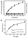
Figure 5. Intrinsic NTPase activity of YchF. (A) 5 μM YchF was incubated with 75 μM [γ-32P]-ATP (●), 15 μM [γ-32P]-GTP (▲), 75 μM [γ-32P]-GTP (■), or 125 μM [γ-32P]-GTP (♦) at 37°C. Samples were taken after different time points and liberated γ-32Pi was extracted and subsequently quantified by scintillation counting. (B) Michaelis-Menten titration of 5 μM YchF and increasing concentrations of [γ-32P]-ATP. Initial rates were plotted against the ATP concentration and fit with a hyperbolic function to yield the catalytic constants vmax of 1.8 ± 0.1 μM min−1 and KM of 41.3 ± 5.8 μM.
None
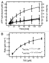
Figure 6. Ribosome-stimulated ATPase activity of YchF. (A) Time courses of ATP hydrolysis of 1 μM YchF and 125 μM [γ-32P]-ATP in the absence (●) and presence of 5 μM 70S (▲), 5 μM 50S (■) or 5 μM 30S (♦) ribosomes. Reactions were incubated at 37°C and 5 μL aliquots were taken at different time points to follow the liberation of γ-32Pi. (B) Michaelis-Menten titration with 70S ribosomes. One μM YchF and 125 μM [γ-32P]-ATP were incubated with increasing concentrations of 70S ribosomes. 70S concentration dependence of the initial rates was fit with the Michaelis-Menten equation to obtain a vmax of 3.1 ± 0.2 μM min−1 and a KM of 7.7 ± 1.1 μM.
None
Figure 7. Minimal Model of YchF function: YchF in its compact ATP-bound state binds to certain “special” 70S ribosomes (70S*). Interaction with 70S* stimulates YchF’s ATPase activity and ATP is hydrolyzed to ADP. In its open ADP-bound state YchF has a lower affinity for 70S* and dissociates. Subsequently, YchF is recharged with ATP and becomes competent for another round of 70S* binding and catalysis.

References

    1. Koonin EV. Comparative genomics, minimal gene-sets and the last universal common ancestor. Nat Rev Microbiol. 2003;1:127–36. doi: 10.1038/nrmicro751. - DOI - PubMed
    1. Wittinghofer A, Vetter IR. Structure-function relationships of the G domain, a canonical switch motif. Annu Rev Biochem. 2011;80:943–71. doi: 10.1146/annurev-biochem-062708-134043. - DOI - PubMed
    1. Caldon CE, March PE. Function of the universally conserved bacterial GTPases. Curr Opin Microbiol. 2003;6:135–9. doi: 10.1016/S1369-5274(03)00037-7. - DOI - PubMed
    1. Mittenhuber G. Comparative genomics of prokaryotic GTP-binding proteins (the Era, Obg, EngA, ThdF (TrmE), YchF and YihA families) and their relationship to eukaryotic GTP-binding proteins (the DRG, ARF, RAB, RAN, RAS and RHO families) J Mol Microbiol Biotechnol. 2001;3:21–35. - PubMed
    1. Galperin MY, Koonin EV. ‘Conserved hypothetical’ proteins: prioritization of targets for experimental study. Nucleic Acids Res. 2004;32:5452–63. doi: 10.1093/nar/gkh885. - DOI - PMC - PubMed

Publication types

MeSH terms

LinkOut - more resources