. 2012 Nov;9(11):1331-8.
doi: 10.4161/rna.22180. Epub 2012 Sep 20.

The human tRNA m (5) C methyltransferase Misu is multisite-specific

Affiliations

The human tRNA m (5) C methyltransferase Misu is multisite-specific

Sylvie Auxilien et al. RNA Biol. 2012 Nov.

Abstract

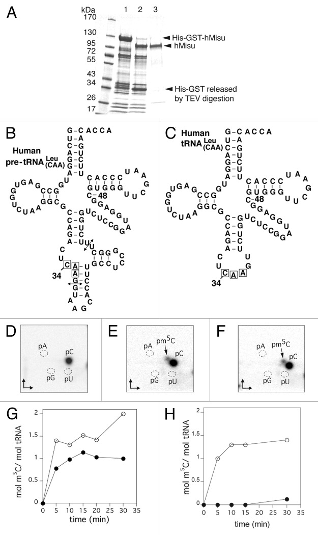
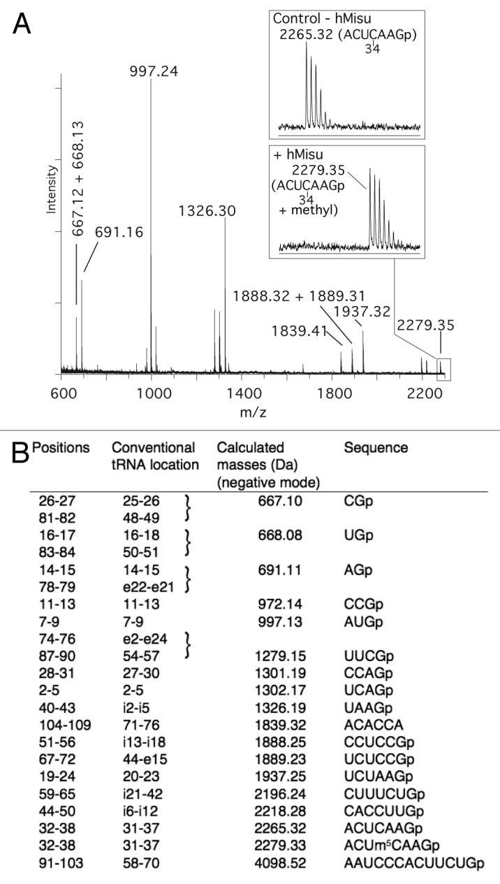
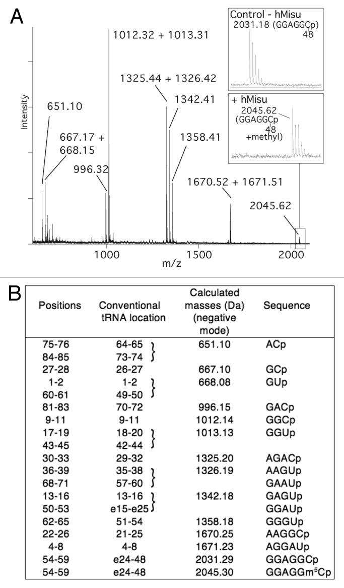
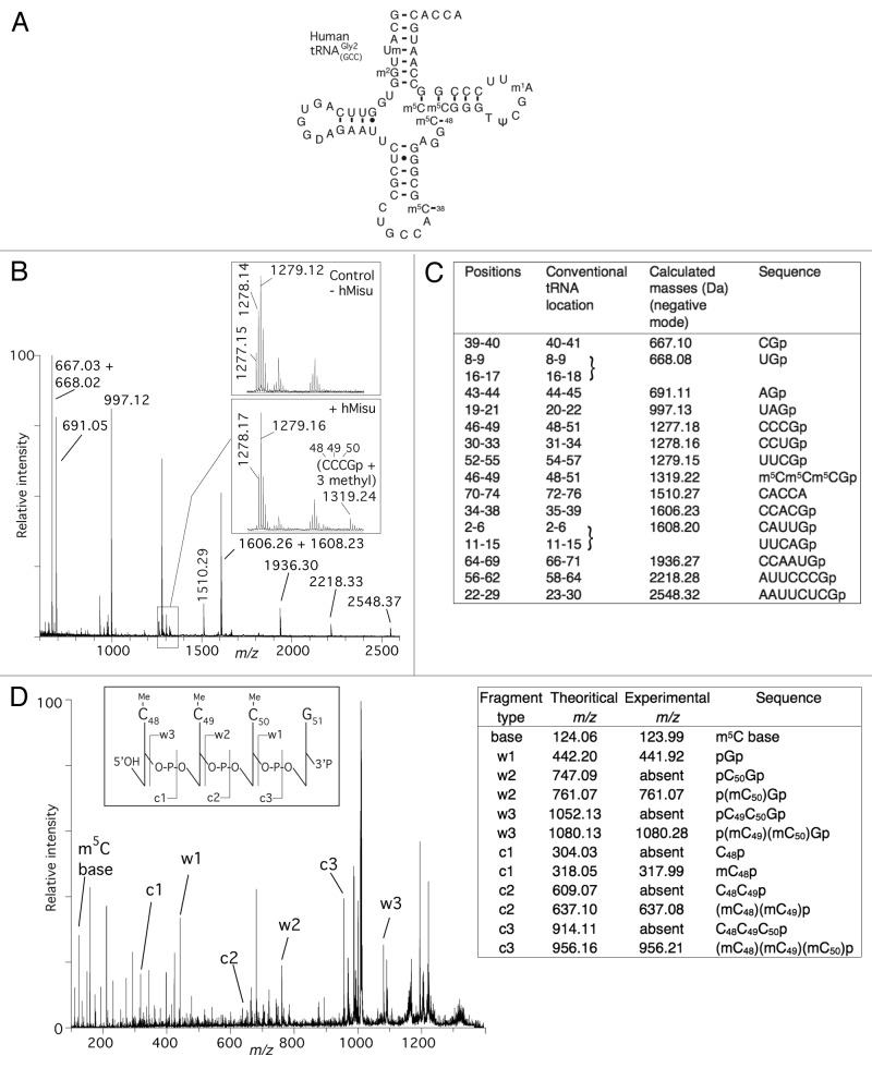
The human tRNA m ( 5) C methyltransferase Misu is a novel downstream target of the proto-oncogene Myc that participates in controlling cell division and proliferation. Misu catalyzes the transfer of a methyl group from S-adenosyl-L-methionine to carbon 5 of cytosines in tRNAs. It was previously shown to catalyze in vitro the intron-dependent formation of m ( 5) C at the first position of the anticodon (position 34) within the human pre-tRNA (Leu) (CAA). In addition, it was recently reported that C48 and C49 are methylated in vivo by Misu. We report here the expression of hMisu in Escherichia coli and its purification to homogeneity. We show that this enzyme methylates position 48 in tRNA (Leu) (CAA) with or without intron and positions 48, 49 and 50 in tRNA (Gly2) (GCC) in vitro. Therefore, hMisu is the enzyme responsible for the methylation of at least four cytosines in human tRNAs. By comparison, the orthologous yeast enzyme Trm4 catalyzes the methylation of carbon 5 of cytosine at positions 34, 40, 48 or 49 depending on the tRNAs.

Keywords: 5-methylcytosine; Misu; NSun2; RNA methyltransferase; Trm4; m5C; tRNA modification enzyme.


Figures

None
Figure 1. Formation of m5C in human tRNA Leu(CAA) catalyzed by purified hMisu. (A) 10% SDS PAGE analysis of hMisu purification. Lane 1: Ni-NTA purified His-GST fused hMisu (117 kDa). Lane 2: Proteins mixture after TEV protease digestion of Ni-NTA purified His-GST-hMisu (after digestion, the molecular weight of hMisu is 86 kDa). Lane 3: hMisu after TEV protease digestion and elution of Ni-NTA column with 20 mM imidazole. (B and C) The transcripts used as substrates were the intron-containing human pre- tRNA Leu(CAA) (B) and intron-less human tRNA Leu(CAA). (C) The arrows indicate the position of the intron. (D−F) Thin layer chromatography analyses of pre- tRNA Leu(CAA) incubated without protein (D) or with hMisu (E), and of tRNA Leu(CAA) incubated with hMisu (F) in the absence of magnesium. The main spot is C and the minor spot m5C. (G and H) Time course of m5C formation catalyzed by hMisu in pre- tRNA Leu(CAA) (G) or tRNA Leu(CAA) (H) in the presence of 50 mM NaCl, and in the absence (open circles) or presence (filled circles) of 10 mM MgCl2.
None
Figure 2. MALDI mass spectrometry analysis of intron-containing human pre- tRNA Leu(CAA) for methylation at position 34. (A) MALDI mass spectrum of pre- tRNA Leu(CAA) methylated by hMisu and digested by RNase T1 that cleaves after guanosines. The spectral region around the ACUCAAGp fragment containing C34 is enlarged to show the peak of the nonmethylated ion (m/z 2265.32) in the control without enzyme and the ion methylated by hMisu (m/z 2279.35). (B) List of theoretical masses of RNase T1 fragments of singly protonated pre- tRNA Leu(CAA).
None
Figure 3. MALDI mass spectrometry analysis of intron-less human tRNA Leu(CAA) for methylation by hMisu at position 48. (A) MALDI mass spectrum of tRNA Leu(CAA) methylated by hMisu and digested by RNase A that cleaves after pyrimidines. The spectral region around the fragment GGAGGCp containing C48 is enlarged to show the peak of the nonmethylated ion (m/z 2031.18) from the control without enzyme and the ion methylated by hMisu (m/z 2045.62). (B) List of theoretical masses of RNase A fragments of singly protonated tRNA Leu(CAA).
None
Figure 4. MALDI mass spectrometry analysis of human tRNA Gly2GCC for methylation by hMisu. (A) Cloverleaf structure of the mature human tRNAGly2GCC indicating its natural posttranscriptional modifications. (B) MALDI mass spectrum of tRNA Gly2GCC methylated by hMisu and digested by RNase T1 that cleaves after guanosines. The spectral region around the fragment CCCGp containing C48, C49 and C50 is enlarged to show the peak of the nonmethylated ion (m/z 1277.15) from the control without enzyme and the ion methylated by hMisu (m/z 1319.24), see also Figure S3. (C) List of theoretical masses of RNase T1 fragments of singly protonated tRNAGly2GCC. (D) Identification of the three methylation targets of hMisu at positions 48, 49 and 50 in human tRNAGly2GCC by MS/MS. The fragment at m/z 1319.24 of the methylated sample shown in (B) was selected and fragmented by tandem mass spectrometry. The fragments assignment follows the scheme of McLuckey et al. The peaks corresponding to the c-ions and w-ions are generated by loss of the 3′ and 5′ nucleotides, respectively.

References

    1. He C. Grand challenge commentary: RNA epigenetics? Nat Chem Biol. 2010;6:863–5. doi: 10.1038/nchembio.482. - DOI - PubMed
    1. Yi C, Pan T. Cellular dynamics of RNA modification. Acc Chem Res. 2011;44:1380–8. doi: 10.1021/ar200057m. - DOI - PMC - PubMed
    1. Nawrot B, Sochacka E, Düchler M. tRNA structural and functional changes induced by oxidative stress. Cell Mol Life Sci. 2011;68:4023–32. doi: 10.1007/s00018-011-0773-8. - DOI - PMC - PubMed
    1. Jenner LB, Demeshkina N, Yusupova G, Yusupov M. Structural aspects of messenger RNA reading frame maintenance by the ribosome. Nat Struct Mol Biol. 2010;17:555–60. doi: 10.1038/nsmb.1790. - DOI - PubMed
    1. Demeshkina N, Jenner L, Yusupova G, Yusupov M. Interactions of the ribosome with mRNA and tRNA. Curr Opin Struct Biol. 2010;20:325–32. doi: 10.1016/j.sbi.2010.03.002. - DOI - PubMed

Publication types

MeSH terms