Review
. 2012 Sep;4(3):129-34.
doi: 10.1038/ijos.2012.59. Epub 2012 Sep 21.

A post-classical theory of enamel biomineralization… and why we need one

Affiliations
Review

A post-classical theory of enamel biomineralization… and why we need one

James P Simmer et al. Int J Oral Sci. 2012 Sep.

Abstract

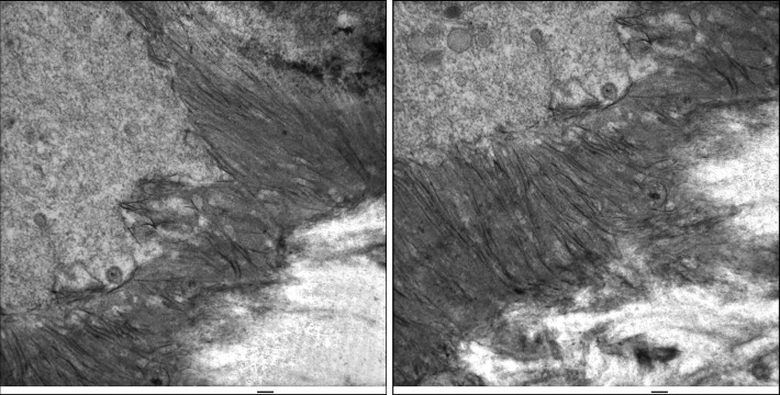
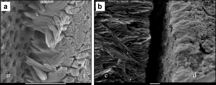
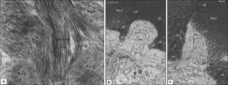
Enamel crystals are unique in shape, orientation and organization. They are hundreds of thousands times longer than they are wide, run parallel to each other, are oriented with respect to the ameloblast membrane at the mineralization front and are organized into rod or interrod enamel. The classical theory of amelogenesis postulates that extracellular matrix proteins shape crystallites by specifically inhibiting ion deposition on the crystal sides, orient them by binding multiple crystallites and establish higher levels of crystal organization. Elements of the classical theory are supported in principle by in vitro studies; however, the classical theory does not explain how enamel forms in vivo. In this review, we describe how amelogenesis is highly integrated with ameloblast cell activities and how the shape, orientation and organization of enamel mineral ribbons are established by a mineralization front apparatus along the secretory surface of the ameloblast cell membrane.


Figures

Figure 1
Figure 1
Formation of initial enamel. Day 7 mouse mandibles were fixed with 2.5% glutaraldehyde in sodium cacodylate buffer and post-fixed with osmium tetroxide. Sections were stained with uranyl acetate, then lead citrate, and viewed by TEM. Ameloblasts are on the upper left. Banded collagen fibers are on the lower right. The enamel ribbons initiate on the dentin surface in close association with collagen and the mineralization front on the ameloblast membrane. Scale bars=100 nm. TEM, transmission electron microscopy.
Figure 2
Figure 2
Preferred fracture levels in Klk4 and Mmp20 null mice. (a) Klk4 null mouse enamel tends to fracture where the initial enamel forms the first interrod enamel (ir) and the base of the rods (r) just above the DEJ. Bar=10 µm. (b) Mmp20 null mouse enamel (e) separates from dentin (d) right at the DEJ. Bar=1 µm. DEJ, dentino-enamel junction; KLK4, kallikrein 4; MMP20, matrix metalloproteinase 20.
Figure 3
Figure 3
Crystal shape, orientation, and organization are determined at the mineralization front. Three TEMs of an undecalcified sections of secretory stage inner enamel stained with uranyl acetate and lead citrate reproduced with permission from (a) Rat incisor section showing that within the rods, crystallites run parallel to the rod axis (×70 000). Between adjacent rods the interrod crystallites run at almost right angles. (b) Developing human tooth showing the relationship of the ameloblasts to the interrod and rod growth regions (×20 000). The Tomes' process (T) is surrounded by interrod enamel (IR) and is lined by a relatively smooth membrane. Interrod growth regions (igr) are seen at the prong tips on either side. The section has cut the rod growth region tangentially and it thus appears in the center of the process (im). dcw: distal cell web. (c) The rod growth region (rgr) and the forming rod are seen on one surface of Tomes' process (T). The opposite surface faces interrod enamel (lR). Interrod growth regions (igr) are seen at the prong tips on either side. Infolded membranes (im) are seen at interrod and rod growth regions. TEM, transmission electron microscopy.

References

    1. Selvig KA. The crystal structure of hydroxyapatite in dental enamel as seen with the electron microscope. J Ultrastruct Res. 1972;41 3:369–375. - PubMed
    1. Meckel AH, Griebstein WJ, Neal RJ. Structure of mature human dental enamel as observed by electron microscopy. Arch Oral Biol. 1965;10 5:775–783. - PubMed
    1. Kerebel B, Daculsi G, Kerebel LM. Ultrastructural studies of enamel crystallites. J Dent Res. 1979;58 Spec Issue B:844–851. - PubMed
    1. Daculsi G, Menanteau J, Kerebel LM, et al. Length and shape of enamel crystals. Calcif Tissue Int. 1984;36 5:550–555. - PubMed
    1. Aoba T. Recent observations on enamel crystal formation during mammalian amelogenesis. Anat Rec. 1996;245 2:208–218. - PubMed

Publication types

Substances