Methods for studying tooth root cementum by light microscopy
- PMID: 22996273
- PMCID: PMC3464984
- DOI: 10.1038/ijos.2012.57
Methods for studying tooth root cementum by light microscopy
Abstract
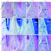
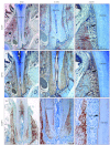
The tooth root cementum is a thin, mineralized tissue covering the root dentin that is present primarily as acellular cementum on the cervical root and cellular cementum covering the apical root. While cementum shares many properties in common with bone and dentin, it is a unique mineralized tissue and acellular cementum is critical for attachment of the tooth to the surrounding periodontal ligament (PDL). Resources for methodologies for hard tissues often overlook cementum and approaches that may be of value for studying this tissue. To address this issue, this report offers detailed methodology, as well as comparisons of several histological and immunohistochemical stains available for imaging the cementum-PDL complex by light microscopy. Notably, the infrequently used Alcian blue stain with nuclear fast red counterstain provided utility in imaging cementum in mouse, porcine and human teeth. While no truly unique extracellular matrix markers have been identified to differentiate cementum from the other hard tissues, immunohistochemistry for detection of bone sialoprotein (BSP), osteopontin (OPN), and dentin matrix protein 1 (DMP1) is a reliable approach for studying both acellular and cellular cementum and providing insight into developmental biology of these tissues. Histological and immunohistochemical approaches provide insight on developmental biology of cementum.
Figures


Similar articles
-
Deficiency in acellular cementum and periodontal attachment in bsp null mice.J Dent Res. 2013 Feb;92(2):166-72. doi: 10.1177/0022034512469026. Epub 2012 Nov 26. J Dent Res. 2013. PMID: 23183644 Free PMC article.
-
Mineralization defects in cementum and craniofacial bone from loss of bone sialoprotein.Bone. 2015 Sep;78:150-64. doi: 10.1016/j.bone.2015.05.007. Epub 2015 May 9. Bone. 2015. PMID: 25963390 Free PMC article.
-
Immunohistochemical study of bone sialoprotein and osteopontin in healthy and diseased root surfaces.J Periodontol. 2006 Oct;77(10):1665-73. doi: 10.1902/jop.2006.060087. J Periodontol. 2006. PMID: 17032108
-
Role of two mineral-associated adhesion molecules, osteopontin and bone sialoprotein, during cementogenesis.Connect Tissue Res. 1995;33(1-3):1-7. doi: 10.3109/03008209509016974. Connect Tissue Res. 1995. PMID: 7554941 Review.
-
Developmental pathways of periodontal tissue regeneration: Developmental diversities of tooth morphogenesis do also map capacity of periodontal tissue regeneration?J Periodontal Res. 2019 Feb;54(1):10-26. doi: 10.1111/jre.12596. Epub 2018 Sep 12. J Periodontal Res. 2019. PMID: 30207395 Review.
Cited by
-
Mechanical Forces Exacerbate Periodontal Defects in Bsp-null Mice.J Dent Res. 2015 Sep;94(9):1276-85. doi: 10.1177/0022034515592581. Epub 2015 Jun 30. J Dent Res. 2015. PMID: 26130257 Free PMC article.
-
Loss of Discoidin Domain Receptor 1 Predisposes Mice to Periodontal Breakdown.J Dent Res. 2019 Dec;98(13):1521-1531. doi: 10.1177/0022034519881136. Epub 2019 Oct 14. J Dent Res. 2019. PMID: 31610730 Free PMC article.
-
Overlapping functions of bone sialoprotein and pyrophosphate regulators in directing cementogenesis.Bone. 2017 Dec;105:134-147. doi: 10.1016/j.bone.2017.08.027. Epub 2017 Sep 1. Bone. 2017. PMID: 28866368 Free PMC article.
-
Global proteome profiling of dental cementum under experimentally-induced apposition.J Proteomics. 2016 Jun 1;141:12-23. doi: 10.1016/j.jprot.2016.03.036. Epub 2016 Apr 16. J Proteomics. 2016. PMID: 27095596 Free PMC article.
-
Development of immortalized Hertwig's epithelial root sheath cell lines for cementum and dentin regeneration.Stem Cell Res Ther. 2019 Jan 3;10(1):3. doi: 10.1186/s13287-018-1106-8. Stem Cell Res Ther. 2019. PMID: 30606270 Free PMC article.
References
-
- Foster B, Popowics T, Fong H, et al. Advances in defining regulators of cementum development and periodontal regeneration. Curr Top Dev Biol. 2007;78:47–126. - PubMed
-
- Diekwisch T. The developmental biology of cementum. Int J Dev Biol. 2001;45 5/6:695–706. - PubMed
-
- Bosshardt D. Are cementoblasts a subpopulation of osteoblasts or a unique phenotype. J Dent Res. 2005;84 5:390–406. - PubMed
-
- Bosshardt D, Selvig K. Dental cementum: the dynamic tissue covering of the root. Periodontol 2000. 1997;13:41–75. - PubMed
-
- Bosshardt D, Schroeder H. Cementogenesis reviewed: a comparison between human premolars and rodent molars. Anat Rec. 1996;245 2:267–292. - PubMed
Publication types
MeSH terms
Substances
Grants and funding
LinkOut - more resources
Full Text Sources
Research Materials

