. 2012 Sep 20;17(9):11216-28.
doi: 10.3390/molecules170911216.

Salvianolic acid A protects the peripheral nerve function in diabetic rats through regulation of the AMPK-PGC1α-Sirt3 axis

Affiliations

Salvianolic acid A protects the peripheral nerve function in diabetic rats through regulation of the AMPK-PGC1α-Sirt3 axis

Xiaoyan Yu et al. Molecules. .

Abstract

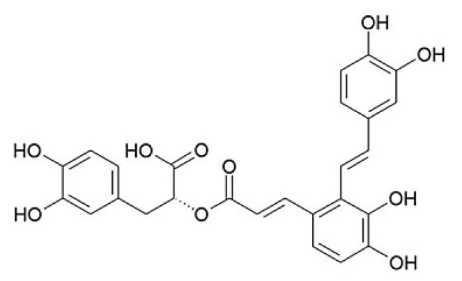
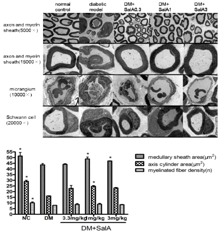
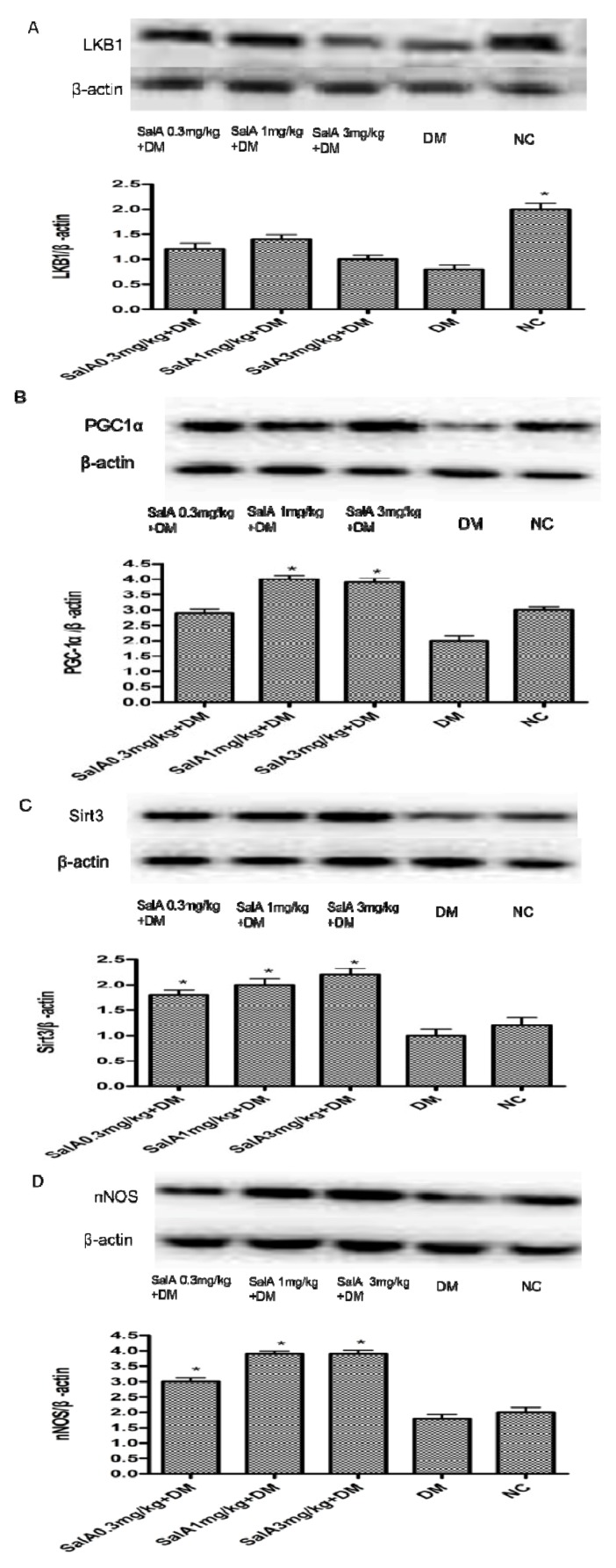
Salvianolic acid A (SalA) is one of the main efficacious, water-soluble constituents of Salvia miltiorrhiza Bunge. This study investigated the protective effects of SalA on peripheral nerve in diabetic rats. Administration of SalA (0.3, 1 and 3 mg/kg, ig) was started from the 5th week after strepotozotocin (STZ60 mg/kg) intraperitoneal injection and continued for 8 weeks. Paw withdrawal mechanical threshold (PWMT) and motor nerve conduction velocity (MNCV) were used to assess peripheral nerve function. The western blot methods were employed to test the expression levels of serine-threonine liver kinase B1 (LKB1), AMP-activated protein kinase (AMPK), peroxisome proliferator-activated receptor-gamma coactivator-1alpha (PGC-1α), silent information regulator protein3 (sirtuin 3/Sirt3) and neuronal nitric oxide synthase (nNOS) in sciatic nerve. Results showed that SalA administration could increase PWMT and MNCV in diabetic rats; reduce the deterioration of sciatic nerve pathology; increase AMPK phosphorylation level, up-regulate PGC-1α, Sirt3 and nNOS expression, but had no influence on LKB1. These results suggest that SalA has protective effects against diabetic neuropathy. The beneficial effects of SalA on peripheral nerve function in diabetic rats might be attributed to improvements in glucose metabolism through regulation of the AMPK-PGC1α-Sirt3 axis.


Figures

Figure 1
Figure 1
Chemical structure of SalA.
Figure 2
Figure 2
Effect of SalA on diabetic rat pain withdrawal threshold (A) and motor nerve conduction velocity (B) after 8-weeks treatment. Values are expressed as the mean ± standard deviation (SD), * p < 0.05 compared with diabetic rats. NC = normal control, DM = diabetic model, n = 12.
Figure 3
Figure 3
Representative electronmicrographs of sciatic nerves in rats treated by SalA. Pictures show the cross section of sciatic nerve fiber (axon and myelin sheath), micrangium in sciatic nerve, schwann cell of sciatic nerve in normal control group, diabetic model group and SalA 0.3, 1, 3 mg/kg-treated group; column show the effect of SalA on quantitative ultrastructural analyses of sciatic nerve in diabetic rats after 8 weeks’ treatment (5,000×). * p < 0.05 vs. diabetic model group, n = 4.
Figure 4
Figure 4
Representative Western blot analyses of p-AMPKβ and AMPKβ (A), p-AMPKα and AMPKα (B) in rat sciatic nerves. Protein expression was analyzed by western blot, and bands were quantified by densitometry. β-Actin was used as a loading control and for relative quantification in all cases. # p < 0.05 compared with diabetic control. * p < 0.05 compared with diabetic control. NC = normal control, DM = diabetic model, n = 6.
Figure 5
Figure 5
Representative Western blot analyses of LKB1, PGC-1α, Sirt3 and nNOS (A, B, C and D) in rat sciatic nerves. Protein expression was analyzed by western blot, and bands were quantified by densitometry. β-actin was used as a loading control and for relative quantification in all cases. * p < 0.05 compared with diabetic control. NC = normal control, DM = diabetic model, n = 6.

Similar articles

Cited by

References

    1. Fernyhough P., Calcutt N.A. Abnormal calcium homeostasis in peripheral neuropathies. Cell Calcium. 2010;47:130–139. doi: 10.1016/j.ceca.2009.11.008. - DOI - PMC - PubMed
    1. Tesfaye S., Boulton A.J., Dyck P.J., Freeman R., Horowitz M., Kempler P., Lauria G., Malik R.A., Spallone V., Vinik A., et al. Diabetic neuropathies: Update on definitions, diagnostic criteria, estimation of severity, and treatments. Diabetes Care. 2010;33:2285–2293. doi: 10.2337/dc10-1303. - DOI - PMC - PubMed
    1. Brownlee M. Biochemistry and molecular cell biology of diabetic complications. Nature. 2001;414:813–820. doi: 10.1038/414813a. - DOI - PubMed
    1. Huang Q., Wu Y.T., Tan H.L., Ong C.N., Shen H.M. A novel function of poly(ADP-ribose) polymerase-1 in modulation of autophagy and necrosis under oxidative stress. Cell Death Differ. 2009;16:264–277. doi: 10.1038/cdd.2008.151. - DOI - PubMed
    1. Ronnett G.V., Ramamurthy S., Kleman A.M., Landree L.E., Aja S. AMPK in the brain: Its roles in energy balance and neuroprotection. J. Neurochem. 2009;109:17–23. doi: 10.1111/j.1471-4159.2009.05916.x. - DOI - PMC - PubMed

Publication types

MeSH terms