

Three-dimensional densitometric analysis of maxillary sutural changes induced by rapid maxillary expansion
- PMID: 22996394
- PMCID: PMC3699014
- DOI: 10.1259/dmfr/71798010
Three-dimensional densitometric analysis of maxillary sutural changes induced by rapid maxillary expansion
Abstract
Objective: This prospective study evaluated the density of the midpalatal and transverse sutures as assessed by low-dose CT before rapid maxillary expansion (T0), at the end of active expansion (T1) and after a retention period of 6 months (T2).
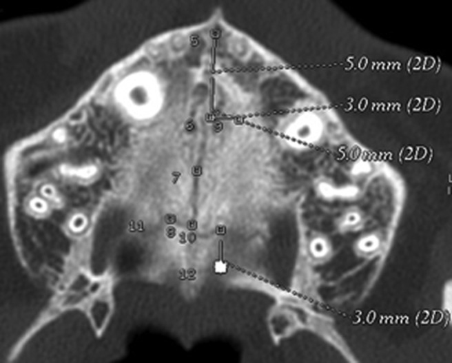
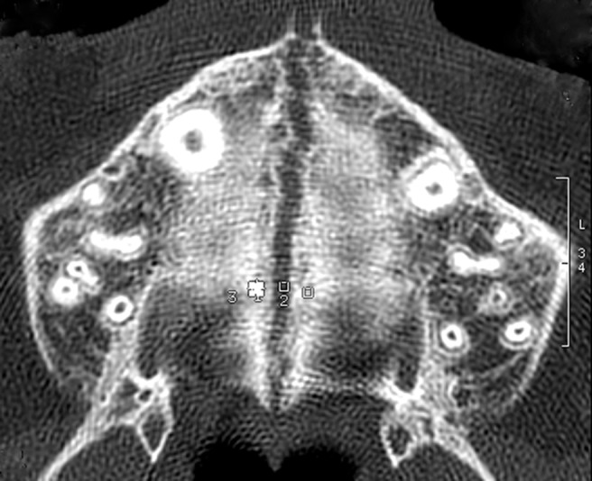
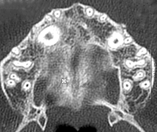
Methods: The study sample comprised 17 pre-pubertal subjects (mean age 11.2 years) with constricted maxillary arches. Total amount of expansion was 7 mm in all subjects. Multislice low-dose CT scans were taken at T0, T1 and T2. On the axial CT scanned images six regions of interest (ROIs) were placed along the midpalatal and transverse sutures and two in maxillary and palatal bony areas. Density was measured in Hounsfield units. Mann-Whitney U test and Friedman analysis of variance with post hoc tests were used (p < 0.05).
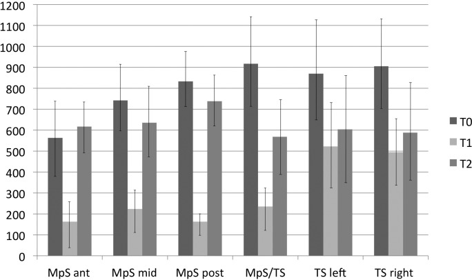
Results: The three ROIs in the midpalatal suture showed a significant decrease in density from T0 to T1, a significant increase from T1 to T2 and a lack of statistically significant differences from T0 to T2. Both ROIs located in the transverse suture showed a significant decrease in density from T0 to T1, followed by a non-significant increase in density from T1 to T2.
Conclusions: At the end of the active phase of expansion a significant reduction in density along the midpalatal and transverse sutures was observed in all subjects. The sutural density of the midpalatal suture at T2 indicated reorganization of the midpalatal suture while the density along the transverse suture increased without reaching the pre-treatment values, possibly due to different morphology between midpalatal and transverse sutures.
Figures
Comment in
-
To beam or not to beam: that is the question.Dentomaxillofac Radiol. 2013;42(2):20120375. doi: 10.1259/dmfr.20120375. Dentomaxillofac Radiol. 2013. PMID: 23393294 Free PMC article. No abstract available.
References
-
- Cameron CG, Franchi L, Baccetti T, McNamara JA., Jr Long-term effects of rapid maxillary expansion: A posteroanterior cephalometric evaluation. Am J Orthod Dentofacial Orthop 2002; 121: 129–135 - PubMed
-
- Haas AJ. Rapid expansion of the maxillary dental arch and nasal cavity by opening the midpalatal suture. Angle Orthod 1961; 31: 73–90
-
- McNamara JA. Maxillary transverse deficiency. Am J Orthod Dentofacial Orthop 2000; 117: 567–570 - PubMed
-
- Melsen B. Palatal growth studied on human autopsy material. A histologic microradiographic study. Am J Orthod 1975; 68: 42–54 - PubMed
-
- Melsen B, Melsen F. The postnatal development of the palatomaxillary region studied on human autopsy material. Am J Orthod 1982; 82: 329–342 - PubMed
MeSH terms
LinkOut - more resources
Full Text Sources
Other Literature Sources
Medical
