. 2012 Oct;122(10):3781-90.
doi: 10.1172/JCI64413. Epub 2012 Sep 4.

Pemphigus autoantibodies generated through somatic mutations target the desmoglein-3 cis-interface

Affiliations

Pemphigus autoantibodies generated through somatic mutations target the desmoglein-3 cis-interface

Giovanni Di Zenzo et al. J Clin Invest. 2012 Oct.

Abstract

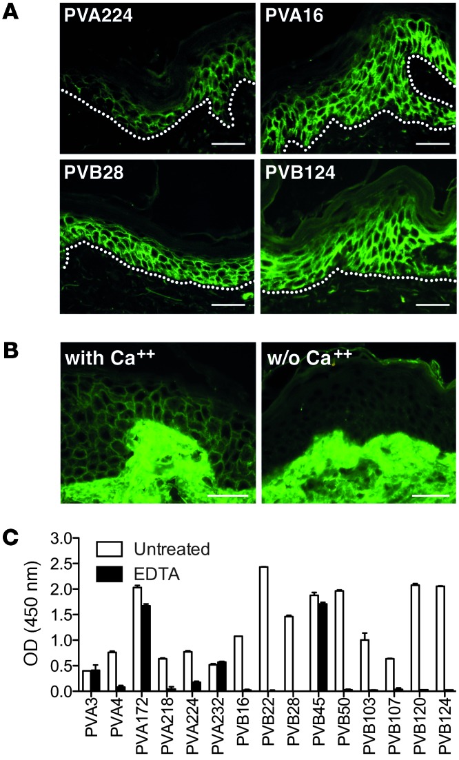
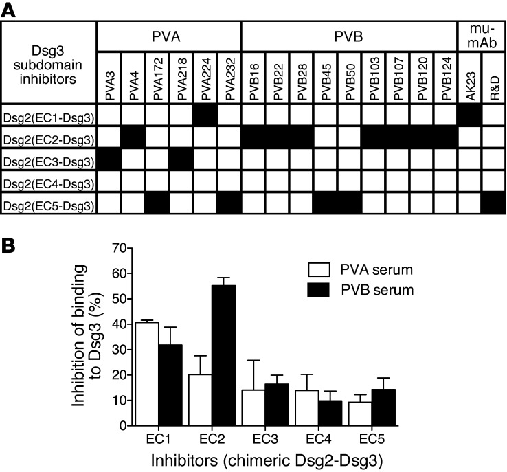
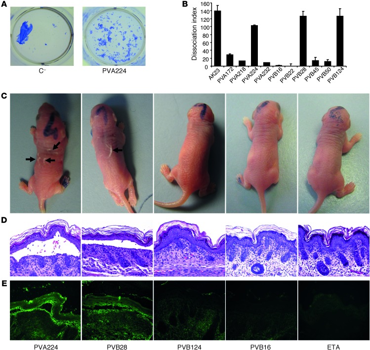
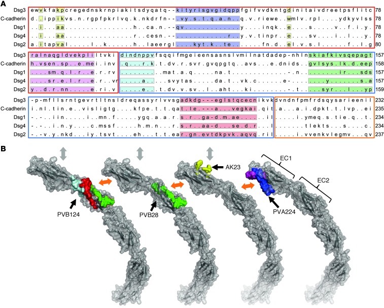
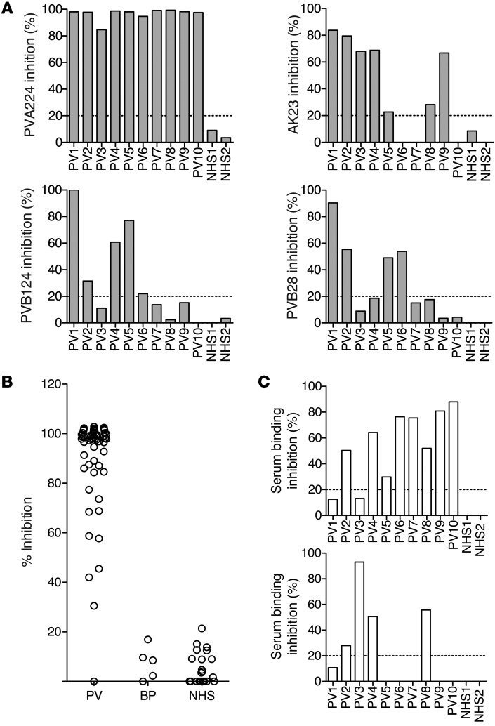
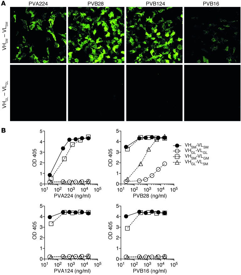
Pemphigus vulgaris (PV) is an autoimmune blistering disease of skin and mucous membranes caused by autoantibodies to the desmoglein (DSG) family proteins DSG3 and DSG1, leading to loss of keratinocyte cell adhesion. To learn more about pathogenic PV autoantibodies, we isolated 15 IgG antibodies specific for DSG3 from 2 PV patients. Three antibodies disrupted keratinocyte monolayers in vitro, and 2 were pathogenic in a passive transfer model in neonatal mice. The epitopes recognized by the pathogenic antibodies were mapped to the DSG3 extracellular 1 (EC1) and EC2 subdomains, regions involved in cis-adhesive interactions. Using a site-specific serological assay, we found that the cis-adhesive interface on EC1 recognized by the pathogenic antibody PVA224 is the primary target of the autoantibodies present in the serum of PV patients. The autoantibodies isolated used different heavy- and light-chain variable region genes and carried high levels of somatic mutations in complementary-determining regions, consistent with antigenic selection. Remarkably, binding to DSG3 was lost when somatic mutations were reverted to the germline sequence. These findings identify the cis-adhesive interface of DSG3 as the immunodominant region targeted by pathogenic antibodies in PV and indicate that autoreactivity relies on somatic mutations generated in the response to an antigen unrelated to DSG3.


Figures

Figure 1
Figure 1. Human antibodies bind conformational epitopes in the DSG3 ectodomain.
(A) Cryosections of human skin were stained with biotinylated human mAbs isolated from PV patients. Shown are 4 representative antibodies out of the 15 analyzed. The cutaneous basement membrane zone is marked by a dotted line. Scale bars: 100 μm. (B) Staining of human epidermis performed as in A with antibody PVB28 in the presence (with) or absence (w/o) of Ca+. Scale bars: 100 μm. (C) Binding of the indicated antibodies to DSG3-coated plates in the presence or absence of EDTA as assessed by ELISA. Data are representative of 2 separate experiments (mean ± SEM).
Figure 2
Figure 2. DSG3 subdomain specificity of DSG3-specific autoantibodies.
(A) Inhibition of mAb binding to coated DSG3 by chimeric molecules consisting of DSG2 carrying a single DSG3 subdomain (EC1, EC2, EC3, EC4, or EC5). Black squares indicate a significant inhibition of antibody binding. Two mouse mAbs (mu-mAb), AK23 and RD, specific for EC1 and EC5, respectively, were also analyzed. Results are representative of 2 independent experiments. (B) Inhibition of PVA and PVB serum antibody binding to coated DSG3 by chimeric DSG2–DSG3 molecules carrying the indicated DSG3 subdomains. Shown are mean values ± SEM of 2 independent experiments.
Figure 3
Figure 3. In vitro and in vivo pathogenic activity of anti-DSG3 antibodies.
(A) Representative keratinocyte dissociation experiment. Primary human keratinocytes were seeded to confluence, and mAb PVA224 and a control antibody were added (1–10 μg/ml), followed by addition of ETA to cleave DSG1 molecules. Cells were incubated with dispase I to detach the monolayer from the plate, and sheet fragments were fixed and stained using crystal violet. (B) Keratinocyte dissociation induced by different antibodies. Shown is the dissociation index determined as specified in Methods in duplicate cultures in 2 independent experiments (mean ± SEM). (CE) Macroscopic blisters (black arrows) (C), intraepidermal suprabasal blistering and acantholysis (D), and intercellular deposition of human IgG in the epidermis (E) induced by injection into newborn mice of the indicated antibodies coadministered with ETA. Data are representative of results obtained from groups of 5 mice. Original magnification, ×200.
Figure 4
Figure 4. Epitope mapping of 3 pathogenic antibodies.
(A) Alignment of DSG3 amino acid sequence 1–232 with C-cadherin, DSG1, DSG2, and DSG4. EC1, EC2, and EC3 subdomains are highlighted with red, blue, and orange boxes, respectively. Peptides recognized by PVA224 are highlighted in blue and pink, peptides recognized by PVB28 in green, and peptides recognized by PVB124 in light blue, red, and green. The reported amino acid residues bound by AK23 (13) are highlighted in yellow. (B) Location of peptides recognized by PVA224, PVB28, PVB124, and AK23 on the structure of C-cadherin ectodomain as determined in ref. . Color code is as above. The trans-adhesive interface is indicated by a gray double arrow. The cis-adhesive interfaces are indicated by orange double arrows.
Figure 5
Figure 5. The PVA224 site on DSG3 EC1 is an immunodominant target for pathogenic autoantibodies in PV patients.
(A) The inhibitory capacity of 10 PV and 2 control sera on the binding of human (PVA224, PVB124, and PVB28) and mouse (AK23) pathogenic antibodies to DSG3. (B) The inhibitory capacity of patient or control sera to inhibit binding of PVA224 to DSG3. Sera from 57 PV patients, 10 bullous pemphigoid (BP) patients, and 32 healthy donors (NHS) are shown. (C) The inhibitory capacity of murinized PVA224 (top) or mouse AK23 (bottom) on the binding of serum antibodies from 10 PV patients and 2 controls. Cut-off value (dashed line) is the mean of inhibition value of the 40 control sera plus 2 SD. Similar results were obtained in 2 independent experiments.
Figure 6
Figure 6. DSG3 autoreactivity is determined by somatic mutations in the VH genes.
Four antibodies were produced recombinantly in the original VH/VL somatically mutated version (VHSM-VLSM), in a full germlined VH/VL version (VHGL-VLGL), or a VH only or VL only germlined version (VHGL-VLSM or VHSM-VLGL, respectively). The antibodies were tested for staining of 293T cells transfected with DSG3 gene (A) and binding to DSG3-coated plates as assessed by ELISA (B). Representative experiment out 3 independent experiments performed. The low binding of the germlined PVB28 was also observed on uncoated plates and can be explained by a low degree of polyspecificity.

References

    1. Amagai M, Klaus-Kovtun V, Stanley JR. Autoantibodies against a novel epithelial cadherin in pemphigus vulgaris, a disease of cell adhesion. Cell. 1991;67(5):869–877. doi: 10.1016/0092-8674(91)90360-B. - DOI - PubMed
    1. Ishii K, et al. Characterization of autoantibodies in pemphigus using antigen-specific enzyme-linked immunosorbent assays with baculovirus-expressed recombinant desmogleins. J Immunol. 1997;159(4):2010–2017. - PubMed
    1. Ruach M, Ohel G, Rahav D, Samueloff A. Pemphigus vulgaris and pregnancy. Obstet Gynecol Surv. 1995;50(10):755–760. doi: 10.1097/00006254-199510000-00023. - DOI - PubMed
    1. Anhalt GJ, et al. Induction of pemphigus in neonatal mice by passive transfer of IgG from patients with the disease. N Engl J Med. 1982;306(20):1189–1196. doi: 10.1056/NEJM198205203062001. - DOI - PubMed
    1. Amagai M, Koch PJ, Nishikawa T, Stanley JR. Pemphigus vulgaris antigen (desmoglein 3) is localized in the lower epidermis, the site of blister formation in patients. J Invest Dermatol. 1996;106(2):351–355. doi: 10.1111/1523-1747.ep12343081. - DOI - PubMed

Publication types

MeSH terms