

The structure of the fruit peel in two varieties of Malus domestica Borkh. (Rosaceae) before and after storage
- PMID: 22996687
- PMCID: PMC3659274
- DOI: 10.1007/s00709-012-0454-y
The structure of the fruit peel in two varieties of Malus domestica Borkh. (Rosaceae) before and after storage
Abstract
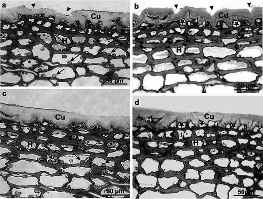
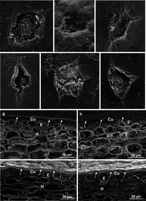
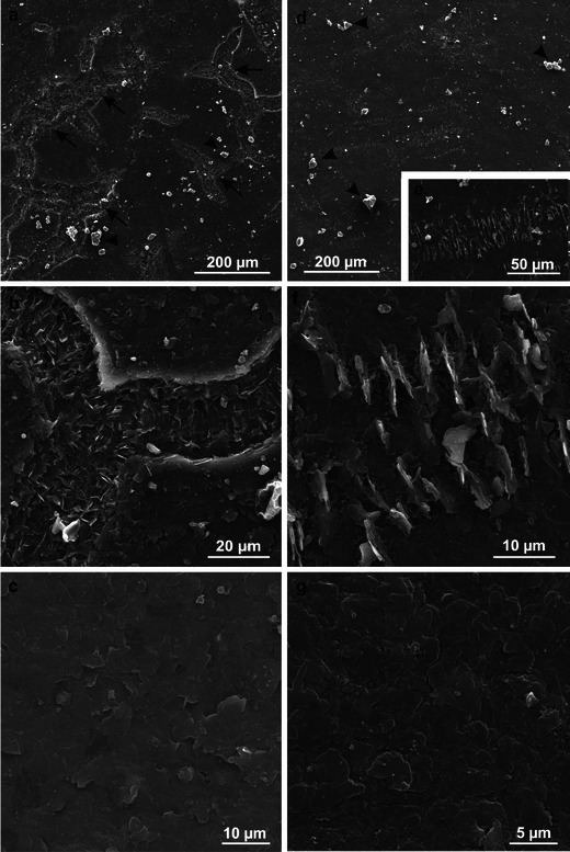
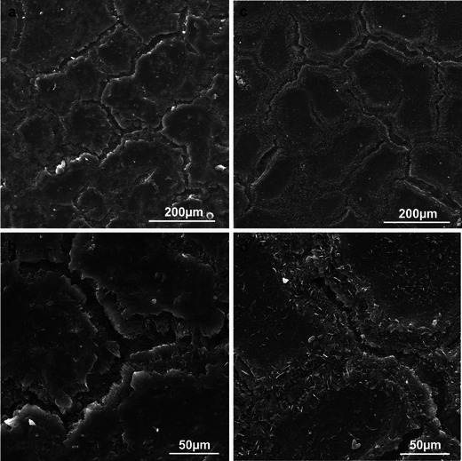
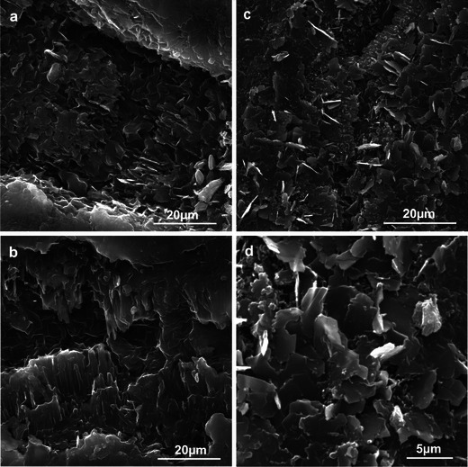
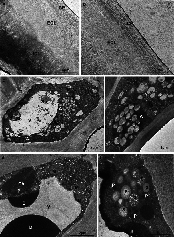
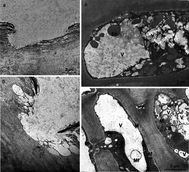
The structure of fruit peel of two apple varieties 'Szampion' and 'Jonagold' was investigated using light microscopy as well as scanning and transmission electron microscopy. The samples were taken immediately after harvest and after 6-month controlled atmosphere storage. The Szampion and Jonagold fruit differed in terms of the surface type, number of lenticels, thickness of the cuticular epithelium, height of epidermal cells and thickness of the hypodermis as well as the amount of crystalline wax and the number of microcracks formed on the fruit surface. The 6-month storage resulted in fruit weight loss, increased numbers and depth of microcracks, thickening of the amorphous wax layer and enhanced production of platelet forms of crystalline wax, which filled the microcracks abundantly. Compared with Jonagold, the Szampion fruit exhibited a fewer lenticels, a bigger number of microcracks, smaller amounts of crystalline wax and more substantial weight loss. The apple varieties studied had a reticulate-lamellate cuticle, and at harvest, the epidermal and hypodermal cells contained numerous amyloplasts filled with starch grains, which were not found after the storage period. Additionally, after storage, the cell protoplasts in the apple peel displayed a disorganised structure, and their vacuoles contained fragments of cell membranes, intravacuolar precipitates and deposits, and spherical bodies. The results may facilitate better understanding of changes occurring in fruits of Szampion and Jonagold during storage and help choose the best storage conditions to reduce loss of weight and prevent impairment of fruit quality.
Figures
References
-
- Babos K, Sass P, Mohácsy P. Relationship between the peel structure and storability of apples. Acta Agron Hung. 1984;33:41–50.
-
- Baker EA. Chemistry and morphology of plant epicuticular waxes. In: Cutler DF, Alvin KL, Price CE, editors. The plant cuticle. London: Academic; 1982. pp. 139–166.
-
- Barthlott W. Scanning electron microscopy of the epidermal surface in plants. In: Claugher D, editor. Scanning electron microscopy in taxonomy and functional morphology. Oxford: Clarendon; 1990. pp. 157–174.
-
- Barthlott W, Neinhuis C, Cutler D, Ditsch F, Meusel I, Theisen I, Wilhelmi H. Classification and terminology of plant epicuticular waxes. Bot J Linn Soc. 1998;126:237–260. doi: 10.1111/j.1095-8339.1998.tb02529.x. - DOI
-
- Barthlott W, Theisen I, Borsch T, Neinhuis C. Epicuticular waxes and vascular plant systematic: integrating micromorphological and chemical data. In: Stuessy TF, Meyer V, Hörandl E, editors. Deep morphology. Toward a renaissance of morphology in plant systematic. Rugell: GANTER Verlag; 2003. pp. 189–206.
MeSH terms
LinkOut - more resources
Full Text Sources
