. 2012 Oct;122(10):3737-41.
doi: 10.1172/JCI63948. Epub 2012 Sep 17.

Hyperthyroid-associated osteoporosis is exacerbated by the loss of TSH signaling

Affiliations

Hyperthyroid-associated osteoporosis is exacerbated by the loss of TSH signaling

Ramkumarie Baliram et al. J Clin Invest. 2012 Oct.

Abstract

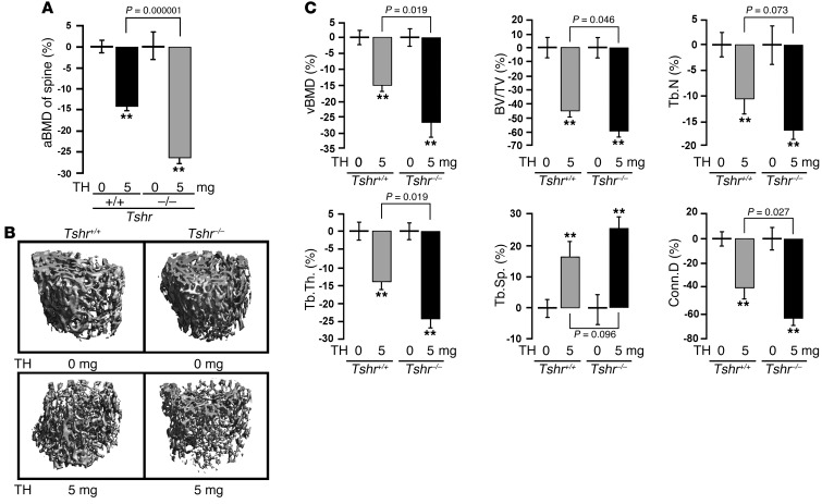
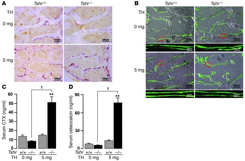
The osteoporosis associated with human hyperthyroidism has traditionally been attributed to elevated thyroid hormone levels. There is evidence, however, that thyroid-stimulating hormone (TSH), which is low in most hyperthyroid states, directly affects the skeleton. Importantly, Tshr-knockout mice are osteopenic. In order to determine whether low TSH levels contribute to bone loss in hyperthyroidism, we compared the skeletal phenotypes of wild-type and Tshr-knockout mice that were rendered hyperthyroid. We found that hyperthyroid mice lacking TSHR had greater bone loss and resorption than hyperthyroid wild-type mice, thereby demonstrating that the absence of TSH signaling contributes to bone loss. Further, we identified a TSH-like factor that may confer osteoprotection. These studies suggest that therapeutic suppression of TSH to very low levels may contribute to bone loss in people.


Figures

Figure 1
Figure 1. Hyperthyroid TSHR-deficient mice have greater bone loss than wild-type hyperthyroid mice with intact TSH signaling.
aBMD (A), micro-CT images (B), and individual parameters from vertebral bodies (C) of wild-type (Tshr+/+) and Tshr–/– mice implanted with 0-mg (placebo) or 5-mg T4 pellets (TH) for 21 days. Mean percent changes (±SEM) in vBMD, BV/TV, Tb.N, Tb.Th, Tb.Sp, and Conn.D are shown. **P < 0.01; n = 6–8 mice/group.
Figure 2
Figure 2. TSHR deficiency accelerates bone remodeling in hyperthyroidism.
Representative images of TRAP-labeled (A) or calcein-labeled bones (B), as well as serum C-telopeptide (CTX) (C) and serum osteocalcin (D) levels in wild-type and Tshr–/– mice implanted with 0-mg (placebo) or 5-mg sustained-release T4 pellets for 21 days. Scale bars in A and B: 100 μm; B inset, 10 μm. **P < 0.01, P < 0.01; n = 4–9 mice/group.

References

    1. Sawin CT, Geller A, Hershman JM, Castelli W, Bacharach P. The aging thyroid. The use of thyroid hormone in older persons. JAMA. 1989;261(18):2653–2655. doi: 10.1001/jama.1989.03420180077034. - DOI - PubMed
    1. Bassett JH, Williams GR. The skeletal phenotypes of TRalpha and TRbeta mutant mice. J Mol Endocrinol. 2009;42(4):269–282. doi: 10.1677/JME-08-0142. - DOI - PubMed
    1. Földes J, Tarján G, Szathmari M, Varga F, Krasznai I, Horvath C. Bone mineral density in patients with endogenous subclinical hyperthyroidism: is thyroid status a risk factor for osteoporosis? Clin Endocrinol (Oxf). 1993;39(5):521–527. doi: 10.1111/j.1365-2265.1993.tb02403.x. - DOI - PubMed
    1. Abe E, et al. TSH is a negative regulator of skeletal remodeling. Cell. 2003;115(2):151–162. doi: 10.1016/S0092-8674(03)00771-2. - DOI - PubMed
    1. Sampath TK, et al. Thyroid-stimulating hormone restores bone volume, microarchitecture, and strength in aged ovariectomized rats. J Bone Miner Res. 2007;22(6):849–859. doi: 10.1359/jbmr.070302. - DOI - PubMed

Publication types

MeSH terms