. 2012 Oct;122(10):3678-91.
doi: 10.1172/JCI63635. Epub 2012 Sep 17.

Myocardin regulates BMP10 expression and is required for heart development

Affiliations

Myocardin regulates BMP10 expression and is required for heart development

Jianhe Huang et al. J Clin Invest. 2012 Oct.

Abstract

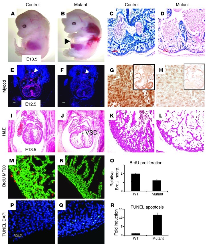
Myocardin is a muscle lineage-restricted transcriptional coactivator that has been shown to transduce extracellular signals to the nucleus required for SMC differentiation. We now report the discovery of a myocardin/BMP10 (where BMP10 indicates bone morphogenetic protein 10) signaling pathway required for cardiac growth, chamber maturation, and embryonic survival. Myocardin-null (Myocd) embryos and embryos harboring a cardiomyocyte-restricted mutation in the Myocd gene exhibited myocardial hypoplasia, defective atrial and ventricular chamber maturation, heart failure, and embryonic lethality. Cardiac hypoplasia was caused by decreased cardiomyocyte proliferation accompanied by a dramatic increase in programmed cell death. Defective chamber maturation and the block in cardiomyocyte proliferation were caused in part by a block in BMP10 signaling. Myocardin transactivated the Bmp10 gene via binding of a serum response factor-myocardin protein complex to a nonconsensus CArG element in the Bmp10 promoter. Expression of p57kip2, a BMP10-regulated cyclin-dependent kinase inhibitor, was induced in Myocd-/- hearts, while BMP10-activated cardiogenic transcription factors, including NKX2.5 and MEF2c, were repressed. Remarkably, when embryonic Myocd-/- hearts were cultured ex vivo in BMP10-conditioned medium, the defects in cardiomyocyte proliferation and p57kip2 expression were rescued. Taken together, these data identify a heretofore undescribed myocardin/BMP10 signaling pathway that regulates cardiomyocyte proliferation and apoptosis in the embryonic heart.


Figures

Figure 1
Figure 1. Myocd–/– embryos exhibit hypoplastic hearts and heart failure.
(A and B) In situ hybridization analysis performed with radiolabeled antisense myocardin riboprobe to sections prepared from E9.5 control (A) and Myocd–/– (B) embryos demonstrating myocardin mRNA (arrowheads, pink signal) throughout the common atria (A) and ventricle (V) of control mice, but only background levels in Myocd–/– hearts. Original magnification, ×40. (C and D) Whole-mount preparation of E9.5 (C) and E10.5 (D) WT (+/+) and Myocd–/– (–/–) mutant mice. E10.5 mutants exhibit growth retardation and pericardial effusion (arrowheads) indicative of heart failure. (EH) Hearts harvested from E10.0 WT (E and F) and Myocd–/– mutant (G and H) embryos shown from frontal (E and G) and dorsal (F and H) perspective, demonstrating that both the WT and mutant hearts have completed the stage of looping morphogenesis. The future right and left atria (RA, LA), right ventricle, left ventricle, and OFT are identified. (IL) Scanning electron micrographs of hearts harvested from E9.5 WT (I and J) and Myocd–/– mutant (K and L) embryos, demonstrating that the Myocd–/– mutant ventricle (L) is hypoplastic and underdeveloped compared with the WT heart (J). Original magnification, ×200 (I and K); ×500 (J and L). See also Supplemental Figure 1 and Supplemental Videos 1–6.
Figure 2
Figure 2. Myocd–/– mutant embryos develop a block in chamber maturation.
(AH) H&E-stained sections of E8.75 WT (AD) and Myocd–/– (EH) embryos demonstrating initiation of looping morphogenesis with identifiable common atria, common ventricle, bulbus-cordis (BC), bulbus-ventricular region (BV), bulbus-truncus (BT), and aortic sac (AS). A single endocardial (EN) layer is observed at this stage of development. The compact zone of the WT ventricle (C and D) is composed of 4–6 layers of densely packed cardiac myocytes (Myo), while the compact zone of Myocd–/– hearts is composed of 1–3 layers of cardiac myocytes. Original magnification, ×200. (IP) At E9.0, the AVC and cardiac OFT are well developed in WT hearts (IL). In Myocd–/– hearts, the AVC and OFT are wide open and cardiac jelly in these regions is substantially decreased (MP). In addition, in Myocd–/– hearts, the compact and trabecular zones of the primitive ventricle are thinned, disorganized, and markedly underdeveloped. Original magnification, ×200. (QX) At E9.5, control (QT) and Myocd–/– mutants (UX) have completed looping morphogenesis and normally undergo chamber maturation. The paired dorsal aortas (arrowheads, Ao) are seen in control and mutant embryos at this stage of development. Compared with control hearts, the compact zone of Myocd–/– mutant hearts is hypocellular (arrowheads) and disorganized, and there is a block in development of trabecular (Trab) myocardium. Original magnification, ×100 (QS and UW); ×400 (T and X).
Figure 3
Figure 3. Myocd–/– hearts exhibit decreased cardiomyocyte proliferation and increased apoptosis.
(AL) Assessment of cardiomyocyte proliferation in hearts of E9.5 WT and Myocd–/– (null) embryos. Histological sections showing hearts of E9.5 WT and Myocd–/– (null) embryos were immunostained with antibodies that recognize pHH3 Ser10 (AC; orange stain), PCNA (DF; green stain), cyclin D2 (GI; green stain), and BrdU (JL; green stain). White bars represent WT and black bars represent Myocd–/– embryos. Immunostained cardiomyocytes in the bulbus-cordis (RV) and future left ventricle (LV) were quantified by a blinded observer, and data are expressed as mean number of immunostained ventricular cardiomyocytes per high power field in Myocd–/– hearts compared with WT hearts ± SEM (P < 0.01, control versus mutant for each marker). (MO) Analysis of apoptosis in E8.5–E9.5 control (WT) and Myocd–/– mutant (null) hearts by TUNEL staining. (MO) Sections of E.8.5, E9.0, and E9.5 WT and Myocd–/– (null) embryos were immunostained for TUNEL activity (green) and costained with DAPI (blue). TUNEL-positive cardiomyocytes per high power field were quantified by a blinded observer, and data are expressed as mean percentage of TUNEL-positive cardiomyocytes ± SEM (P < 0.01, control versus mutant for each marker). (PS) EM of E9.5 WT and Myocd–/– mutant hearts revealed nuclear chromatin aggregation, nuclear fragmentation, and cytoplasmic apoptotic body formation (Figure 2Q, arrowheads) in hearts of Myocd–/– embryos. Mitochondrial swelling with breakdown of the mitochondrial membranes was also observed in Myocd–/– hearts (Figure 2S, arrowheads). Original magnification, ×50,000.
Figure 4
Figure 4. Nkx2-5Cre+/MyocdF/F mutant embryos develop hypoplastic hearts attributable to a block in cell proliferation and increased apoptosis.
(A and B) E13.5 Nkx2-5Cre+/MyocdF/F mutant embryo (B) exhibits generalized edema and pericardial effusions (arrowhead) compared with MyocdF/F control embryo (A). (C and D) E10.5 control Nkx2-5Cre+/MyocdF/+/Rosa26 (C) and Nkx2-5Cre+MyocdF/F/Rosa 26 embryos (D) demonstrating intense blue staining (LacZ) throughout the myocardium; scale bars: 100 μm. (E and F) In situ hybridization analyses demonstrating myocardin mRNA (pink signal) throughout control hearts (E) and background levels in mutant hearts (F). Comparable myocardin is observed in the trachea and esophagus (arrowheads). Original magnification, ×40. (G and H) E12.5 MyocdF/F control (G) and Nkx2-5Cre+/MyocdF/F mutant (H) embryos demonstrating nuclear expression of myocardin (dark brown stain) in control hearts (G) and attenuated expression in mutant hearts. Inset panel shows low-power magnification of heart and surrounding tissues. Original magnification, ×400; ×40 (inset). (IL) E13.5 MyocdF/F control (I and K) and Nkx2-5Cre+/MyocdF/F mutant (J and L) embryos demonstrating thinning of the compact zone and trabecular myocardium and VSD (arrow) in the mutant hearts. Original magnification, ×40 (I and J); ×200 (K and L). (MO) E13.5 control (M) and conditional mutant (N) embryos immunostained for BrdU (orange) and MF20 (green) expression. Data expressed as relative number of BrdU-expressing cardiomyocytes in the mutant heart compared with the WT heart ± SEM (P < 0.01, control versus mutant). Original magnification, ×400. (PR) Apoptosis quantified in E12.5 control (P) and Nkx2-5Cre+/MyocdF/F (Q) hearts by TUNEL (green stain). Data are expressed as fold induction in TUNEL-positive cardiomyocytes per high power field in mutant compared with control hearts ± SEM (P < 0.01). Original magnification, ×200.
Figure 5
Figure 5. Myocardin regulates expression of key cardiogenic transcription factors and Bmp10 coincident with ventricular chamber maturation.
(A) qRT-PCR was performed with mRNA harvested from the hearts of 3 E9.5 WT control and 3 Myocd–/– mutant embryos as described in Methods. White bars (controls) and black bars (mutants) show the relative level (arbitrary units) of Myocd, Mkl1, Mkl2, Gata4, Srf, Nkx-2.5, Mef2c, Tbx5, and Bmp10 gene expression. Data are expressed as relative gene expression in mutant compared with WT control hearts (arbitrary units) ± SEM. (B) Immunohistochemical analyses of markers of cardiomyocyte differentiation in E9.5 WT control and Myocd–/– (mutant) hearts. Sections were immunostained with antibodies that recognize Gata4, Nkx-2.5, Mef2c, Srf, myosin heavy chain (MF20) (green stain), Mlc2v (red stain), cardiac troponin T (cTnT) (green stain), SMA (red stain), and SM22α (SM22). In situ hybridization analysis performed with radiolabeled antisense Tbx5 riboprobe (red signal) to sections prepared from E9.5 control and Myocd–/– embryos. See also Supplemental Figure 2.
Figure 6
Figure 6. Myocardin regulates Bmp10 signaling and p57kip2 expression in the embryonic heart.
(AD) Sections of E9.0 WT (A and C) and Myocd–/– (B and D) mutant hearts were immunostained with anti-Bmp10 antibody (green stain) and counterstained with DAPI (C and D; blue stain). Bmp10 immunostaining is markedly reduced in Myocd–/– hearts compared with control hearts. (E) Immunoblot analysis of protein lysates prepared from the hearts of E9.5 WT and Myocd–/– embryos. Blots were hybridized to Myocd, Bmp10, Gata4, and β-tubulin antibodies, respectively. Arrows denote pro-BMP and processed Bmp10 protein, respectively. Quantitative analysis of the hybridization signal (normalized to β-tubulin) revealed a 90% decrease in Bmp10 expression in Myocd–/– hearts compared with controls. MW markers are shown to the right of each blot. See also Supplemental Uncut gels. (FI) Sections of E9.5 WT (F and H) and Myocd–/– (G and I) hearts were immunostained with anti-pSmad1/5/8 antibody and counterstained with DAPI (H and I). pSmad1/5/8 expression is markedly reduced in Myocd–/– hearts compared with WT hearts. (JM) Sections of E9.0 WT (J and L) and Myocd–/– (K and M) hearts were immunostained with anti-p57kip2 antibody and costained with eosin. p57kip2 expression is markedly increased in the compact and trabecular zone myocardium of Myocd–/– hearts compared with WT hearts.
Figure 7
Figure 7. Myocardin binds to and transactivates the Bmp10 promoter.
(A) Schematic representation of the Bmp10 promoter and CArG boxes identified (genomic location) in the mouse, rat, human, and chicken BMP10 promoters, respectively. The nucleotide sequence of the mutant CArG box utilized in EMSA and transient transfection analyses is shown below. (B) EMSAs performed with biotin-labeled mouse Bmp10 CArG box oligonucleotide and nuclear extracts prepared from Cos7 cells transfected with an expression plasmid-encoding myocardin, SRF, or pcDNA3. Where indicated, binding reaction mixtures included unlabeled competitor oligonucleotide or anti-SRF antibody. The low-mobility nuclear protein complex containing SRF and myocardin is indicated with a double arrow to the right, and the supershifted complex is denoted by a single arrow. (C) Transient transfection analysis of Cos7 cells cotransfected with pcDNA.MyocdL or the pcDNA.MyocdS expression plasmid and the indicated luciferase reporter plasmid. Luciferase activity is reported as mean luciferase activity compared with luciferase activity in cells cotransfected with the pcDNA3 plasmid ± SEM. (D) ChIP assays performed with chromatin harvested from the hearts of E9.5 embryos. Chromatin was immunoprecipitated with anti-SRF antibody, anti-myocardin antibody, or preimmune rabbit sera (negative control). qRT-PCR was performed with PCR primers flanking either the mouse c-fos promoter CArG box or the mouse Bmp10 promoter CArG box. The upper panels show amplified DNA products immunoprecipitated with either anti-SRF antibody or anti-myocardin antibody. The graph shows qPCR quantification of amplified DNA expressed as fold-enrichment (arbitrary units) normalized to input DNA ± SEM.
Figure 8
Figure 8. Bmp10 rescues the defect in cardiomyocyte proliferation and p57kip2 expression observed in Myocd–/– hearts.
E9.5 WT and Myocd–/– mutant hearts were grown for 48 hours in conditioned medium harvested from NIH3T3 cells that were stably transduced with lentivirus encoding EGFP (control) or mouse Bmp10 (+ Bmp10) (n = 5 hearts per group). (AC) Whole mount of WT heart (A), Myocd–/– heart grown in control medium (B), and Myocd–/– heart grown in Bmp10-conditioned medium (C). Myocd–/– hearts were smaller than WT hearts and beat more slowly and less vigorously. See also Supplemental Videos 7–10. (DF) DAPI staining (blue nuclear stain) of WT, Myocd–/–, and Myocd–/– + Bmp10 explanted hearts. Scale bars in microns are shown. (GI) BrdU incorporation in WT, Myocd–/–, and Myocd–/– explanted hearts grown in Bmp10-conditioned medium. Sections of explanted hearts were immunostained with BrdU antibody (orange stain). BrdU-positive cardiomyocytes per high power field were quantified by a blinded observer, and data are expressed as mean percentage of BrdU-positive cardiomyocytes ± SEM. (JL) pHH3 expression (orange stain) in WT, Myocd–/–, and Myocd–/– explanted hearts grown in Bmp10 conditioned medium. Cardiomyocytes undergoing mitosis in prophase/metaphase exhibited centronuclear staining, while cardiomyocytes in anaphase (arrowheads) exhibit speckled nuclear staining. pHH3-positive cardiomyocytes per high power field were quantified by a blinded observer and data expressed as mean percentage of pHH3-positive cardiomyocytes ± SEM. (MO) Sections immunostained for p57kip2 (brown stain) demonstrating marked increased in p57kip2 in Myocd–/– heart, which decreases significantly when grown in Bmp10-conditioned medium.
Figure 9
Figure 9. Myocardin transduces signals regulating Bmp10 signaling required for cardiac chamber maturation.
In the schematic model, following cardiac looping at the onset of rapid heart cardiac growth associated with atrial and ventricular chamber maturation, myocardin transduces signals that promote binding of a myocardin-SRF protein complex to the nonconsensus CArG box regulating transcription of the Bmp10 gene as well as other CArG boxes regulating transcription of a subset of cardiac-restricted genes. Bmp10, in turn, represses p57kip2, promoting cardiomyocyte proliferation. In addition, Bmp10 is required for maintenance of a subset of key cardiogenic factors, including Nkx2-5 and Mef2c, that regulate structural organization of the cardiomyocyte. Finally, myocardin blocks programmed cell death in the cardiomyocyte, and myocardin deficiency is associated with a dramatic increase in cardiomyocyte apoptosis.

References

    1. Olson EN. A genetic blueprint for growth and development of the heart. Harvey Lect. 2002;98:41–64. - PubMed
    1. Srivastava D, Olson EN. A genetic blueprint for cardiac development. Nature. 2000;407(6801):221–226. doi: 10.1038/35025190. - DOI - PubMed
    1. Wolf M, Basson CT. The molecular genetics of congenital heart disease: a review of recent developments. Curr Opin Cardiol. 2010;25(3):192–197. doi: 10.1097/HCO.0b013e328337b4ce. - DOI - PMC - PubMed
    1. Moon A. Mouse models of congenital cardiovascular disease. Curr Top Dev Biol. 2008;84:171–248. - PubMed
    1. Olson EN. Development. The path to the heart and the road not taken. Science. 2001;291(5512):2327–2328. doi: 10.1126/science.1060063. - DOI - PubMed

Publication types

MeSH terms