. 2012 Nov 9;338(6108):798-802.
doi: 10.1126/science.1219969. Epub 2012 Sep 20.

Germ cell migration across Sertoli cell tight junctions

Affiliations

Germ cell migration across Sertoli cell tight junctions

Benjamin E Smith et al. Science. .

Abstract

The blood-testis barrier includes strands of tight junctions between somatic Sertoli cells that restricts solutes from crossing the paracellular space, creating a microenvironment within seminiferous tubules and providing immune privilege to meiotic and postmeiotic cells. Large cysts of germ cells transit the Sertoli cell tight junctions (SCTJs) without compromising their integrity. We used confocal microscopy to visualize SCTJ components during germ cell cyst migration across the SCTJs. Cysts become enclosed within a network of transient compartments fully bounded by old and new tight junctions. Dissolution of the old tight junctions releases the germ cells into the adluminal compartment, thus completing transit across the blood-testis barrier. Claudin 3, a tight junction protein, is transiently incorporated into new tight junctions and then replaced by claudin 11.


Figures

Fig. 1
Fig. 1. Imaging tight junction fibrils between Sertoli cells
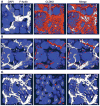
A) The schematic on the left illustrates the orientation of the confocal microscope with respect to the seminiferous tubule. The area imaged is demarcated with a rectangle. The schematic on the right depicts a region of the epithelium prior to preleptotene migration showing germ cells at different steps of differentiation along with supporting Sertoli and peritubular myoid cells. B) Confocal image of SCTJs visualized during Stage IV, a stage in which germ cells are not transiting the SCTJs, using two different markers, F-actin and CLDN11. Bundles of F-actin, labeled with phalloidin, were detected at both the SCTJs and the apical ectoplasmic specializations surrounding the heads of elongating spermatids (*). CLDN11 localized to just the SCTJs. The merged image shows that both markers label the SCTJs. C) An artist’s rendition of the confocal images shown in (B), fig. S2 and movie S1, illustrating three Sertoli cells, the CLDN11-containing tight junctions circumscribing each Sertoli cell and chains of preleptotene spermatocytes connected by intercellular bridges.
Fig. 2
Fig. 2. Formation of transient compartments bounded by tight junctions during migration of preleptotene spermatocytes across Sertoli cell tight junctions
A) During germ cell migration across the SCTJs, Stage IX of the seminiferous epithelium cycle, the leptotene spermatocytes are enclosed within compartments (*) bounded by SCTJs. A double-layer tight junction that sandwiches the leptotene spermatocytes forms the compartments. The arrowhead shows connections between tight junction layers formed at the tricellular junction of three Sertoli cells (S). TEX14 (red) marks the cytoplasmic bridges between the leptotene spermatocytes. TEX14 localization to the tricellular junctions (arrowhead) reveals that the cytoplasmic bridges between spermatocytes pass through the tricellular junctions. B) A schematic of (A) illustrating germ cell migration across the SCTJs showing that the leptotene spermatocytes are transiently enclosed within a network of compartments bounded by tight junctions both apically and basally.
Fig 3
Fig 3. CLDN3 localizes to the newly forming SCTJs
A) Just prior to the migration of preleptotene spermatocytes across the SCTJs, in stage VII of the seminiferous epithelium cycle, CLDN3 (orange) is initially localized to the basal surface of the Sertoli cells (S) rather than the SCTJs (white), labeled with phalloidin. B) In Stage IX of the seminiferous epithelium cycle, when the leptotene spermatocytes migrate across the SCTJs, the spermatocytes are enclosed within the SCTJs (white). However, in this image the basal side of these compartments is incomplete as visualized as breaks in the tight junctions (yellow arrowheads). CLDN3 (orange) localizes to these breaks in the incomplete tight junction fibrils. C) During stage XI of the seminiferous epithelium cycle, after the zygotene spermatocytes (Z) have emerged into the adluminal compartment, CLDN3 (orange) has been almost completely removed from the SCTJs (white). By stage XI, much of the old (apical) tight junctions have been removed.
Fig 4
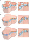
Fig 4. Model for tight junction remodeling during germ cell cyst transit across the blood-testis barrier
A) Schematic illustration of the confocal image in fig. 3A. B) Drawing representing the confocal image in fig. 3B showing segmental localization of CLDN3 (red) and CLDN11 (brown) in areas of new tight junction formation. CLDN11 is usually found in and near tricellular junctions. Once the preleptotene spermatocyte leaves the basement membrane, Stage VIII, the basal surface of the Sertoli cells intercalates between the basement membrane and the preleptotene spermatocyte, forming new Sertoli-Sertoli cell contacts. CLDN3 (red) then localizes to this new contact and forms a new tight junction that encloses the preleptotene spermatocytes. Inset on right shows passage of an intercellular bridge, marked by TEX14 (purple) through a tricellular junction connecting two compartments. C) Illustration of confocal image in fig. 3C showing replacement of CLDN3 with CLDN11 in the new tight junction and removal of the old tight junction.

References

    1. Tsukita S, Furuse M, Itoh M. Multifunctional strands in tight junctions. Nat Rev Mol Cell Biol. 2001 Apr;2:285. - PubMed
    1. Schneeberger EE, Lynch RD. The tight junction: a multifunctional complex. Am J Physiol Cell Physiol. 2004 Jun;286:C1213. - PubMed
    1. Tsukita S, Yamazaki Y, Katsuno T, Tamura A. Tight junction-based epithelial microenvironment and cell proliferation. Oncogene. 2008 Nov 24;27:6930. - PubMed
    1. Dym M, Cavicchia JC. Further observations on the blood-testis barrier in monkeys. Biol Reprod. 1977 Oct;17:390. - PubMed
    1. Weber JE, Russell LD. A study of intercellular bridges during spermatogenesis in the rat. Am J Anat. 1987 Sep;180:1. - PubMed

Publication types

MeSH terms