. 2013 Jan;25(1):61-9.e7.
doi: 10.1111/nmo.12015. Epub 2012 Sep 23.

Nestin-expressing cells in the gut give rise to enteric neurons and glial cells

Affiliations

Nestin-expressing cells in the gut give rise to enteric neurons and glial cells

J Belkind-Gerson et al. Neurogastroenterol Motil. 2013 Jan.

Abstract

Background: Neuronal stem cells (NSCs) are promising for neurointestinal disease therapy. Although NSCs have been isolated from intestinal musclularis, their presence in mucosa has not been well described. Mucosa-derived NSCs are accessible endoscopically and could be used autologously. Brain-derived Nestin-positive NSCs are important in endogenous repair and plasticity. The aim was to isolate and characterize mucosa-derived NSCs, determine their relationship to Nestin-expressing cells and to demonstrate their capacity to produce neuroglial networks in vitro and in vivo.

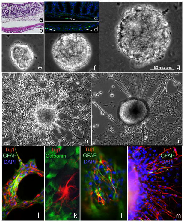
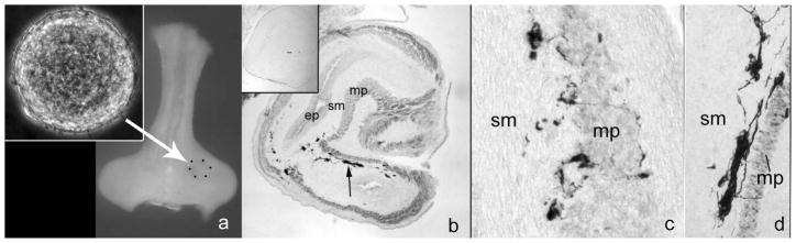
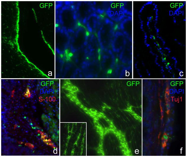
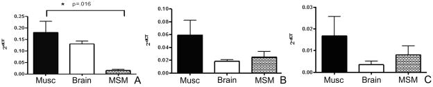
Methods: Neurospheres were generated from periventricular brain, colonic muscularis (Musc), and mucosa-submucosa (MSM) of mice expressing green fluorescent protein (GFP) controlled by the Nestin promoter (Nestin-GFP). Neuronal stem cells were also grown as adherent colonies from intestinal mucosal organoids. Their differentiation potential was assessed using immunohistochemistry using glial and neuronal markers. Brain and gut-derived neurospheres were transplanted into explants of chick embryonic aneural hindgut to determine their fate.

Key results: Musc- and MSM-derived neurospheres expressed Nestin and gave rise to cells of neuronal, glial, and mesenchymal lineage. Although Nestin expression in tissue was mostly limited to glia co-labelled with glial fibrillary acid protein (GFAP), neurosphere-derived neurons and glia both expressed Nestin in vitro, suggesting that Nestin+/GFAP+ glial cells may give rise to new neurons. Moreover, following transplantation into aneural colon, brain- and gut-derived NSCs were able to differentiate into neurons.

Conclusions & inferences: Nestin-expressing intestinal NSCs cells give rise to neurospheres, differentiate into neuronal, glial, and mesenchymal lineages in vitro, generate neurons in vivo and can be isolated from mucosa. Further studies are needed for exploring their potential for treating neuropathies.


Conflict of interest statement

Conflicts of Interest:

No competing interests declared

Figures

Figure 1
Figure 1. Brain and gut neurospheres contain neurons and glia cells
Postnatal mouse colon was separated into MSM (a) and Musc (b) layers, shown by H&E staining. Hu-labeled (green) neuronal cell bodies are present in both layers (c, d, arrows). NS were derived from MSM (e), Musc(f), and brain (g). Scale bar in (g) refers to panels e–g. Following 14 days in culture, adherent brain-derived (h) and Musc-derived (i) neurospheres produce elaborate neuroglial networks. Neuronal (Tuj1) and glial cells (GFAP) are present in Musc-derived NS cultures (j), as are smooth muscle cells (k; calponin). MSM-derived NS (l) similarly contain glial cells (GFAP) and neurons (Tuj1), with neurons extending out from the NS (m).
Figure 2
Figure 2. Transplanted gut-derived neurospheres create neuronal networks in aneural colon
A gut-derived NS (a; inset) was transplanted into aneural E5 chick hindgut (a) and cultured on a CAM for 7 days. Tuj1-immunoreactivity (arrow) was present in the transplanted hindgut (b). No labeling was observed using CN, a chick-specific neuronal antibody, confirming that neurons were mouse-derived (b; inset). Clusters of Tuj1+ cells formed ganglion-like structures extending fine neurites (c, d). ep, epithelium; sm, submucosa; mp, muscularis propria
Figure 3
Figure 3. Nestin is expressed by glial cells in the enteric ganglia
Nestin is strongly expressed in the periventricular region of Nestin-GFP mouse brain (a). In the small intestinal mucosa, GFP+ cells are present around crypts (b) and within the villus core (c). GFP is expressed by cells in submucosal (d) and myenteric (d–f) ganglia. Nestin- immunoreactive cells (GFP+) in the enteric ganglia co-express S-100 (d) but not Tuj1 (f).
Figure 4
Figure 4. Nestin-expressing gut-derived NS co-express neuronal and glial markers
All gut-derived NS are strongly GFP+ (a), and extend intricate neuroglial networks (b–d). Many GFP+ cells also co-express neuronal (c) and glial (d) markers.
Figure 5
Figure 5. Intestinal organoid cultures produce neuroglial colonies
Gut organoid cultures generate colonies that extend long cell processes emerging from their core (a) and form networks with a neural morphology (b). These neuron-like cells were GFP+ (c, d) and, over the following 14 days, proliferate and expand to form larger networks of elongated cells expressing neural (e–g) and glial markers (h).
Figure 6
Figure 6. Markers of pluripotency are expressed in Musc, MSM, and brain neurospheres
Quantitative PCR on proliferating neurospheres demonstrates Sox2 (A), cMyc (B), and Klf4 (C) expression in neurospheres derived from Musc, MSM, and brain. Results represent averages following three separate experiments, each performed in triplicate. The only statistical difference observed was in Sox2 expression between Musc- and MSM-derived neurospheres.

References

    1. Kiba T. Relationships between the autonomic nervous system, humoral factors and immune functions in the intestine. Digestion. 2006;74(3–4):215–27. - PubMed
    1. Margolis KG, Stevanovic K, Karamooz N, et al. Enteric neuronal density contributes to the severity of intestinal inflammation. Gastroenterology. 2011;141(2):588–98. 598 e1–2. - PMC - PubMed
    1. Bischoff SC, Mailer R, Pabst O, et al. Role of serotonin in intestinal inflammation: knockout of serotonin reuptake transporter exacerbates 2,4,6-trinitrobenzene sulfonic acid colitis in mice. Am J Physiol Gastrointest Liver Physiol. 2009;296(3):G685–95. - PubMed
    1. Jessen KR, Mirsky R. Glial cells in the enteric nervous system contain glial fibrillary acidic protein. Nature. 1980;286(5774):736–7. - PubMed
    1. Jessen KR, Mirsky R. Astrocyte-like glia in the peripheral nervous system: an immunohistochemical study of enteric glia. J Neurosci. 1983;3(11):2206–18. - PMC - PubMed

MeSH terms

LinkOut - more resources