. 2012 Sep 21:9:224.
doi: 10.1186/1742-2094-9-224.

Immunoglobulin G (IgG) attenuates neuroinflammation and improves neurobehavioral recovery after cervical spinal cord injury

Affiliations

Immunoglobulin G (IgG) attenuates neuroinflammation and improves neurobehavioral recovery after cervical spinal cord injury

Dung Hoang Nguyen et al. J Neuroinflammation. .

Abstract

Background: Evidence suggests that the inflammatory events in the acute phase of spinal cord injury (SCI) exacerbate the initial trauma to the cord leading to poor functional recovery. As a result, minimizing the detrimental aspects of the inflammatory response after SCI is a promising treatment strategy. In this regard, immunoglobulin G (IgG) from pooled human serum is a promising treatment candidate. Due to its putative, though poorly characterized immuno-modulatory effects, IgG has been used clinically to treat neuroinflammatory disorders such as Guillain-Barré syndrome, but its effects in neurotrauma remain largely unexplored.

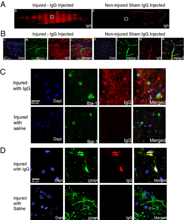
Methods: This study examines the potential neuroprotective effects of IgG in a well-characterized cervical model of SCI. Female Wistar rats were subject to moderate-severe clip compression injury at the C7-T1 level. IgG (0.4 g/kg) or saline was injected intravenously to randomly selected animals at 15 min post SCI. At several time points post SCI, biochemical assays, histology and immunohistochemistry analyses, and neurobehavioral assessments were used to examine the neuroprotective effects of IgG at the molecular, cellular, and neurobehavioral levels.

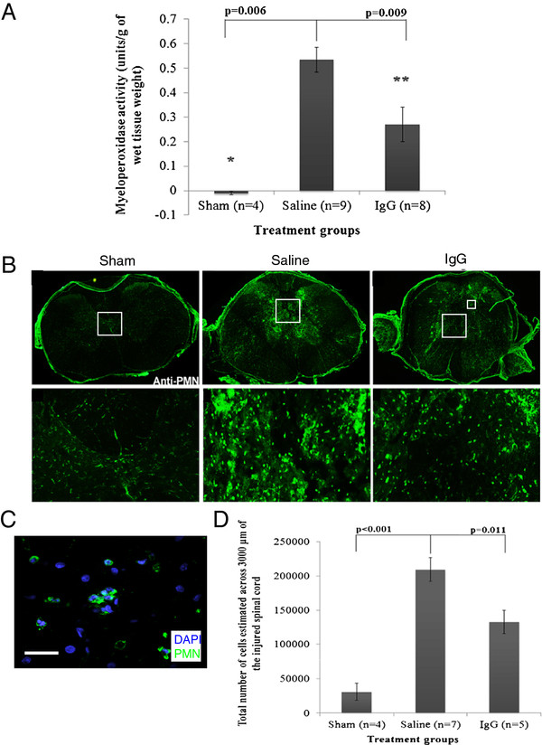
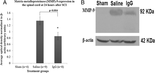
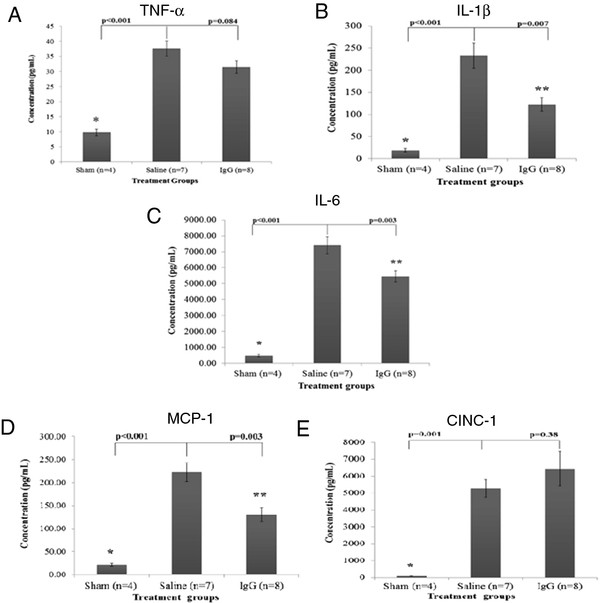
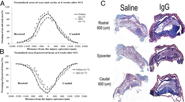
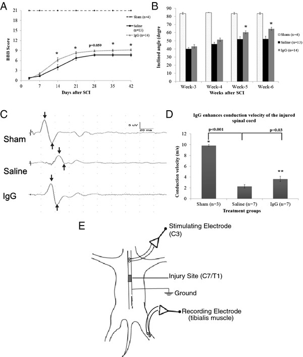
Results: We found that intravenous treatment of IgG following acute clip-compression SCI at C7-T1 significantly reduced two important inflammatory cytokines: interleukin (IL)-1β and IL-6. This early reduction in pro-inflammatory signaling was associated with significant reductions in neutrophils in the spinal cord and reductions in the expression of myeloperoxidase and matrix metalloproteinase-9 in the injured spinal cord at 24 h after SCI. These beneficial effects of IgG were associated with enhanced tissue preservation, improved neurobehavioral recovery as measured by the BBB and inclined plane tests, and enhanced electrophysiological evidence of central axonal conduction as determined by motor-evoked potentials.

Conclusion: The findings from this study indicate that IgG is a novel immuno-modulatory therapy which shows promise as a potential treatment for SCI.


Figures

Figure 1
Figure 1
IgG crosses the blood spinal cord barrier and associates with astrocytes. (A) Representative fluorescence images showing the presence of IgG in the spinal cords of an injured rat (left) and a non-injured rat (right). (B) Vessels were stained with RECA-1 (green) and IgG was labeled with a human specific IgG secondary antibody (red). Confocal images demonstrate IgG was able to cross the blood-spinal cord barrier in injured animals (left panel) but not in non-injured animals (right panel). (C) Representative confocal images of microglia/macrophages (Iba-1; green) and IgG (red). IgG was observed in the parenchyma surrounding cells marked by Iba-1 and DAPI (blue). Although IgG was in the vicinity of Iba-1 positive cells, co-localization between IgG and Iba-1 was not observed. (D) Representative confocal images of astrocytes (GFAP; green) and IgG (red). IgG was observed in the parenchyma surrounding and in the cell soma of GFAP positive astrocytes. The co-localization of IgG and astrocytes suggests potential interaction between IgG and astrocytes. Note that an IgG-positive signal was not observed in the spinal cord of rats injected with saline. Confocal images (B-D) were taken from the boxed area, and scale bars represent 50 μm in length.
Figure 2
Figure 2
IgG reduces neutrophil extravasation. (A) Myeloperoxidase (MPO) activity was detected in sham (n = 4), intravenous saline (saline, n = 9), and intravenous IgG (IgG, n = 8) groups. MPO was used to indirectly measure the presence of neutrophils 24 h after SCI. IgG-treatment significantly reduced MPO activity relative to saline control (ANOVA P <0.05, Bonferroni post-hoc test P = 0.009). (B) Representative fluorescence images demonstrate neutrophil distribution at the injury epicenter in the spinal cord at 24 h post SCI (PMN; green). Neutrophils were observed in both saline and IgG-treated animals. (C) A representative confocal image of neutrophils (PMN; green) and DAPI (blue) was taken from the indicated area (smaller box), and the scale bar represents 75 μm in length. (D) Stereological cell counts demonstrated significantly fewer neutrophils in IgG-treated animals relative to saline control injected animals across 3,000 μm of the injured spinal cord (one-way ANOVA P <0.001; Bonferroni post-hoc test P = 0.011). Error bars represent SEM.
Figure 3
Figure 3
IgG reduces MMP-9 expression. (A) Matrix metalloproteinase-9 (MMP-9) expression was determined by western blot analysis at 24 h after SCI. Three treatment groups were studied including laminectomy only (sham, n = 4), intravenous saline (saline, n = 9), and intravenous IgG (IgG, n = 8). IgG treatment was associated with a significant reduction in MMP-9 level in the injured spinal cord (P = 0.004). Error bars represent SEM. (B) A representative western-blot of MMP-9 is shown. β-actin was used as a loading control.
Figure 4
Figure 4
IgG reduces IL-1β, IL-6, and MCP-1 expression. Expression of cytokines (TNF-α, IL-1β and IL-6) and chemokines (MCP-1 and CINC-1) at 4 h after SCI was determined with ELISA in the spinal cord homogenates of sham, saline, and IgG-treated animals. SCI resulted in a significant increase of all inflammatory mediators relative to non-injured sham animals. (A) There was no difference in TNF-α protein expression between saline and IgG-treated animals (P = 0.084). (B) IgG treatment was associated with a significant reduction in IL-1β following SCI in rats (P = 0.007). (C) IL-6 protein expression was significantly reduced in IgG-treated animals relative to saline controls (P = 0.003). (D) IgG treatment was associated with a significant decrease in MCP-1 protein expression relative to saline controls (P = 0.003). (E) IgG treatment had no effect on the level of CINC-1 after SCI relative to saline control injected animals (P = 0.38). Error bars represent SEM.
Figure 5
Figure 5
IgG reduces scar and cavity formation and preserves neural tissue. (A) The distribution of scar and cavity area over 4000 μm of the injured spinal cords of sham (n = 4), saline (n = 7), and IgG (n = 7) animals was calculated at 6 weeks following injury. There was a treatment effect in the area of cavitation and scar in IgG-treated animals (two-way ANOVA, P = 0.013). IgG-treated animals had significantly less scar and cavity area at 600 μm (P = 0.012) and 720 μm (P = 0.042). (B) Preserved tissue (remaining grey and white matter) in injured spinal cords was calculated. There was an overall treatment effect in the amount of tissue sparing in IgG-treated animals (two-way ANOVA, P = 0.015). IgG-treated animals had significantly more preserved tissue compared to saline-treated animals at a distance of 600 μm (P = 0.002), 720 μm (P = 0.017), and 840 μm (P = 0.044) rostral to the injury epicenter. (C) Representative H&E/LFB images are shown. Error bars represent SEM.
Figure 6
Figure 6
IgG increases neurobehavioral recovery and axonal function. (A) Hind-limb functional recovery was assessed weekly for 6 weeks following SCI. Scores were assigned according to the Basso Beattie Bresnahan (BBB) Scale. Sham animals (n = 4) displayed no hind-limb functional deficits and were given a score of 21. IgG treatment was associated with significant improvement in hind-limb functional recovery (two-way ANOVA P <0.001). The differences in BBB scores between IgG- and saline-treated animals were statistically significant at weeks 2, 3, 5, and 6 (Bonferroni post-hoc test P <0.001, P = 0.015, P = 0.031, and P = 0.017, respectively). (B) The inclined plane test was used to assess functional recovery of the saline (n = 13) and IgG (n = 14) groups. The inclined plane test measures the hind- and forelimb strength and coordination necessary to maintain a horizontal position on an inclined plane. Larger inclined angles are associated with better functional recovery. At weeks 5 and 6, IgG-treated animals performed significantly better than saline-treated animals on the inclined plane test (two-way ANOVA P <0.001; Bonferroni post-hoc test week 5 P = 0.012 and week 6 P <0.001). (C) Motor-evoked potentials (MEPs) were recorded in injured spinal cords at 6 weeks following SCI. Conduction velocity in both the IgG and vehicle control group was significantly reduced compared to uninjured sham rats (one-way ANOVA P <0.05; Bonferroni post-hoc test P <0.01). IgG-treated animals demonstrated significantly faster conduction velocities compared to saline control animals (P = 0.03). (E) Schematic diagram showing how MEPs was recorded in the injured spinal cords at 6 weeks following SCI.

Similar articles

Cited by

References

    1. Hall ED, Yonkers PA, Horan KL. Correlation between attenuation of posttraumatic spinal cord ischemia and preservation of tissue vitamin E by the 21-aminosteroid u74006F: evidence for an in vivo antioxidant mechanism. J Neurotrauma. 1989;6:169–176. doi: 10.1089/neu.1989.6.169. - DOI - PubMed
    1. Tator CH, Fehlings MG. Review of the secondary injury theory of acute spinal cord trauma with emphasis on vascular mechanisms. J Neurosurg. 1991;75:15–26. doi: 10.3171/jns.1991.75.1.0015. - DOI - PubMed
    1. Kwon BK, Sekhon LH, Fehlings MG. Emerging repair, regeneration, and translational research advances for spinal cord injury. Spine. 2010;Suppl. 1:S263–S270. - PubMed
    1. Blight AR. Delayed demyelination, macrophage invasion: a candidate for “secondary” cell damage in spinal cord injury. Cent Nerv Syst Trauma. 1985;2:299–315. - PubMed
    1. Young W. Secondary injury mechanisms in acute spinal cord injury. J Emerg Med. 1993;11:13–22. - PubMed

Publication types

MeSH terms