. 2012 Sep 22:7:70.
doi: 10.1186/1750-1172-7-70.

Reduced elastogenesis: a clue to the arteriosclerosis and emphysematous changes in Schimke immuno-osseous dysplasia?

Affiliations

Reduced elastogenesis: a clue to the arteriosclerosis and emphysematous changes in Schimke immuno-osseous dysplasia?

Marie Morimoto et al. Orphanet J Rare Dis. .

Abstract

Background: Arteriosclerosis and emphysema develop in individuals with Schimke immuno-osseous dysplasia (SIOD), a multisystem disorder caused by biallelic mutations in SMARCAL1 (SWI/SNF-related, matrix-associated, actin-dependent regulator of chromatin, subfamily a-like 1). However, the mechanism by which the vascular and pulmonary disease arises in SIOD remains unknown.

Methods: We reviewed the records of 65 patients with SMARCAL1 mutations. Molecular and immunohistochemical analyses were conducted on autopsy tissue from 4 SIOD patients.

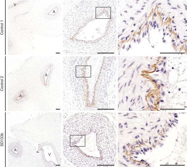
Results: Thirty-two of 63 patients had signs of arteriosclerosis and 3 of 51 had signs of emphysema. The arteriosclerosis was characterized by intimal and medial hyperplasia, smooth muscle cell hyperplasia and fragmented and disorganized elastin fibers, and the pulmonary disease was characterized by panlobular enlargement of air spaces. Consistent with a cell autonomous disorder, SMARCAL1 was expressed in arterial and lung tissue, and both the aorta and lung of SIOD patients had reduced expression of elastin and alterations in the expression of regulators of elastin gene expression.

Conclusions: This first comprehensive study of the vascular and pulmonary complications of SIOD shows that these commonly cause morbidity and mortality and might arise from impaired elastogenesis. Additionally, the effect of SMARCAL1 deficiency on elastin expression provides a model for understanding other features of SIOD.


Figures

Figure 1
Figure 1
Emphysematous lung changes in an SIOD patient. (A, B) Consecutive axial computerized tomography images of the chest of patient SD16 at age 34 years. The images were captured 1 mm apart. Note the lung blebs (arrows).
Figure 2
Figure 2
Photomicrographs of Verhoeff van Gieson stained aortas from SIOD patients and age-matched controls. Compared to age-matched controls, note the decreased elastin fiber staining, the fragmentation and splitting of the elastin fibers, the marked hyperplasia of the tunica intima and the tunica media in the aorta from three individuals with SIOD. Arteries are oriented with the tunica adventitia on the left and the tunica intima on the right; the age of death is in parentheses. Scale bars: 50 μm.
Figure 3
Figure 3
Elastin expression analysis of the umbilical cord from SIOD and unaffected fetuses at 15-weeks gestation. Note that the immunohistochemical analysis shows marked discontinuity and reduced expression of elastin in the internal elastic lamina in SD133b compared to that of 2 age-matched controls. This difference in expression of elastin precedes the development of hypertension, hypercholesterolemia, and renal disease. Abbreviations: A, artery; V, vein. Scale bars: 50 μm.
Figure 4
Figure 4
SMARCAL1 mRNA and protein are expressed in arterial and pulmonary tissue. (A-C) Photomicrographs of immunohistochemical detection of SMARCAL1 in the aorta, common iliac and pulmonary arteries. (D) Photograph of an immunoblot showing expression of SMARCAL1 in aortic smooth muscle cells (AoSMCs), human iliac artery endothelial cells (HIAECs) and aortic adventitial fibroblasts (AoAFs). (E) Photograph of an agarose gel of RT-PCR products showing expression of SMARCAL1 mRNA and cell-specific markers in AoSMCs, HIAECs, and AoAFs. Note that smooth muscle actin (ACTA2) is a marker of myofibroblasts and smooth muscle cells; VE-cadherin (CDH5) is a marker of endothelial cells; and prolyl 4-hydroxylase (P4HA3) is expressed in fibroblasts as well as multiple other cell types [59]. (F-H) Photomicrographs showing immunofluorescent localization of SMARCAL1 (red) and α-tubulin (green) in cultured AoSMCs, HIAECs, and AoAFs. (I-K) Photomicrographs showing immunofluorescent localization of SMARCAL1 (red) and the cell-specific markers smooth muscle actin (I), VE-cadherin (J), and prolyl 4-hydroxylase (K) in AoSMCs, HIAECs, and AoAFs (green), respectively. (L) Photomicrograph of immunohistochemical detection of SMARCAL1 in the lung. (M) Photograph of an immunoblot showing SMARCAL1 expression in normal human lung fibroblasts (NHLFs). (N) Photograph of an agarose gel of RT-PCR products showing expression of SMARCAL1 mRNA and cell-specific markers in NHLFs. GAPDH was used as a control. (O) Photomicrographs showing immunofluorescent localization of SMARCAL1 (red) and α-tubulin (green) in cultured NHLFs. (P) Photomicrographs showing immunofluorescent localization of SMARCAL1 (red) and prolyl 4-hydroxylase (green) and in NHLFs. Scale bars: (A-C, L) 50 μm, 25 μm for inset; (F-K, O, P) 10 μm.
Figure 5
Figure 5
Elastin expression is significantly decreased in the aorta and lung of an SIOD patient. (A) Volcano plot comparing expression of atherosclerosis-related genes in the aorta of SD120 to control aorta. Note the markedly reduced expression of elastin (ELN). Solid grey lines: 4-fold change; solid black line: no change; dotted line: p = 0.01. Grey dots depict genes with decreased expression and black dots depict those with increased expression. (B) Relative elastin protein in the aortic wall of SD120 compared to control. Total elastin protein was measured with the Fastin Elastin Assay. Error bars represent one standard deviation. (C) Plot showing the level of ELN mRNA in SD120 and control lung tissue measured by qRT-PCR. The mRNA levels were standardized to GAPDH mRNA levels and plotted relative to the control. Note the markedly decreased ELN expression. Error bars represent one standard deviation. ** = p < 0.01.
Figure 6
Figure 6
Expression of known ELN transcriptional activators and repressors in patient tissues. (A, B) Plots showing the relative mRNA levels of known ELN transcriptional activators and repressors in SD120 aorta tissue (A) and in SD120 lung tissue (B) compared to controls. The mRNA levels of three independent replicates were standardized to GAPDH mRNA levels and plotted relative to the control. Error bars represent one standard deviation. Abbreviations: NS, not significant; * = p < 0.05, ** = p < 0.01.

References

    1. Clewing JM, Antalfy BC, Lücke T, Najafian B, Marwedel KM, Hori A, Powel RM, Safo Do AF, Najera L, SantaCruz K. et al.Schimke immuno-osseous dysplasia: a clinicopathological correlation. J Med Genet. 2007;44:122–130. - PMC - PubMed
    1. Lücke T, Marwedel KM, Kanzelmeyer NK, Hori A, Offner G, Kreipe HH, Ehrich JH, Das AM. Generalized atherosclerosis sparing the transplanted kidney in Schimke disease. Pediatr Nephrol. 2004;19:672–675. doi: 10.1007/s00467-004-1426-z. - DOI - PubMed
    1. Ehrich JH, Burchert W, Schirg E, Krull F, Offner G, Hoyer PF, Brodehl J. Steroid resistant nephrotic syndrome associated with spondyloepiphyseal dysplasia, transient ischemic attacks and lymphopenia. Clin Nephrol. 1995;43:89–95. - PubMed
    1. Baradaran-Heravi A, Thiel C, Rauch A, Zenker M, Boerkoel CF, Kaitila I. Clinical and genetic distinction of Schimke immuno-osseous dysplasia and cartilage-hair hypoplasia. Am J Med Genet A. 2008;146A:2013–2017. doi: 10.1002/ajmg.a.32406. - DOI - PMC - PubMed
    1. Schimke RN, Horton WA, King CR. Chondroitin-6-sulphaturia, defective cellular immunity, and nephrotic syndrome. Lancet. 1971;2:1088–1089. - PubMed

Publication types

MeSH terms

Supplementary concepts