. 2012 Nov 12;13(11):3850-7.
doi: 10.1021/bm3013023. Epub 2012 Oct 8.

Intravenous hemostatic nanoparticles increase survival following blunt trauma injury

Affiliations

Intravenous hemostatic nanoparticles increase survival following blunt trauma injury

Andrew J Shoffstall et al. Biomacromolecules. .

Abstract

Trauma is the leading cause of death for people ages 1-44, with blood loss comprising 60-70% of mortality in the absence of lethal CNS or cardiac injury. Immediate intervention is critical to improving chances of survival. While there are several products to control bleeding for external and compressible wounds, including pressure dressings, tourniquets, or topical materials (e.g., QuikClot, HemCon), there are no products that can be administered in the field for internal bleeding. There is a tremendous unmet need for a hemostatic agent to address internal bleeding in the field. We have developed hemostatic nanoparticles (GRGDS-NPs) that reduce bleeding times by ~50% in a rat femoral artery injury model. Here, we investigated their impact on survival following administration in a lethal liver resection injury in rats. Administration of these hemostatic nanoparticles reduced blood loss following the liver injury and dramatically and significantly increased 1 h survival from 40 and 47% in controls (inactive nanoparticles and saline, respectively) to 80%. Furthermore, we saw no complications following administration of these nanoparticles. We further characterized the nanoparticles' effect on clotting time (CT) and maximum clot firmness (MCF) using rotational thromboelastometry (ROTEM), a clinical measurement of whole-blood coagulation. Clotting time is significantly reduced, with no change in MCF. Administration of these hemostatic nanoparticles after massive trauma may help staunch bleeding and improve survival in the critical window following injury, and this could fundamentally change trauma care.


Figures

Figure 1
Figure 1
Nanoparticle Schematic and Characterization. a) Hemostatic nanoparticles (GRGDS-NPs) consist of PLGA-PLL biodegradable polymer cores, with PEG arms that expose the GRGDS moiety for targeting activated platelets. b) SEM shows nanoparticle size distribution and morphology. c) 1H-NMR spectral analysis confirms the pegylation of the co-block-polymer and the PEG-coronal structure of the nanoparticles. Deuterated water (top, gray overlay) and deuterated chloroform (bottom, black overlay). d) These are administered intravenously via the tail vein after a partial hepatectomy in the rat. The medial lobe (ML) is transected in this model. Right (RL), left (LL) and caudate lobes (CL) are labeled for reference.
Figure 2
Figure 2
Survival and Blood Loss. a-b) Survival is significantly increased by treatment with the hemostatic GRGDS-functionalized nanoparticles. c-d) The liver mass is tightly controlled in this injury model and is extremely reproducible in size, both in ratio to body mass (1.00% +/− 0.13% S.D.) and in ratio to the remaining liver (22.8 %+/− 2.8% S.D.). The dotted lines on the resected liver graph show the inclusion criteria for this study. e) There is a trend toward a reduction in blood loss with the GRGDS-NP group, but is not significantly significant. f) 100% of animals with a blood volume loss less than 32% survive, but rapidly increases above this threshold. Error bars represent SEM.
Figure 3
Figure 3
Injury Surface Characterization. a) Hemostatic nanoparticles loaded with C6 (green) are found integrated with the adherent clot after it is removed and examined under fluorescent microscopy. b) The majority of bleeding appears to occur from the 2-4 major transected blood vessels in this injury model. c) Scanning electron microscopy is used to verify the presence of the nanoparticles (black arrow) and their integration with the fibrin mesh.
Figure 4
Figure 4
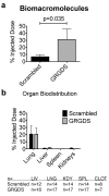
Biodistribution. a) 31% of hemostatic GRGDS-NPs locate in the clot, versus 7% for the scrambled-NP control group. Total mean nanoparticle recovery is 53.7% of total injected dose for GRGDS and 29.6% for scrambled. b) The “Liver” group is representative of the particle distribution to the uninjured lower left lobe of the liver. Minimal particle distribution is found in the spleen and kidneys. Approx. 20% is found in the lungs for each formulation. This may be indicative of microemboli in the lung or nanoparticles still in pulmonary circulation.
Figure 5
Figure 5
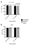
In vitro Testing. Outcomes include CT+CFT (a) and MCF (b). The GRGDS group had a lower (faster) clotting time and a higher clot firmness compared to saline. Error bars represent SEM. CT = Clotting Time; CFT = Clot Formation Time; MCF = Maximum Clot Formation; CT+CFT is the sum total of the time it takes from initiation of the experiment until a 20 mm clot is formed.

References

    1. Kauvar DS, Lefering R, Wade CE. J Trauma. 2006;60(6 Suppl):S3–11. - PubMed
    1. Champion HR, Bellamy RF, Roberts CP, Leppaniemi A. J Trauma. 2003;54(5 Suppl):S13–9. - PubMed
    1. Holcomb JB, McMullin NR, Pearse L, Caruso J, Wade CE, Oetjen-Gerdes L, Champion HR, Lawnick M, Farr W, Rodriguez S, Butler FK. Ann Surg. 2007;245(6):986–91. - PMC - PubMed
    1. Kauvar DS, Wade CE. Crit Care. 2005;9(Suppl 5):S1–9. - PMC - PubMed
    1. Clifford CC. Mil Med. 2004;169(12 Suppl):8–10. 4. - PubMed

Publication types

Substances