. 2012 Sep 20;75(6):1094-104.
doi: 10.1016/j.neuron.2012.08.032.

Metabolic communication between astrocytes and neurons via bicarbonate-responsive soluble adenylyl cyclase

Affiliations

Metabolic communication between astrocytes and neurons via bicarbonate-responsive soluble adenylyl cyclase

Hyun B Choi et al. Neuron. .

Abstract

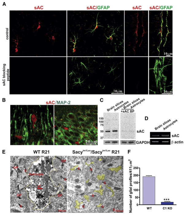
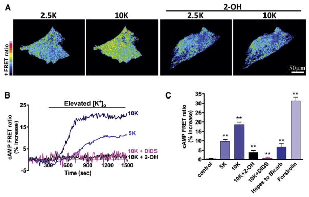
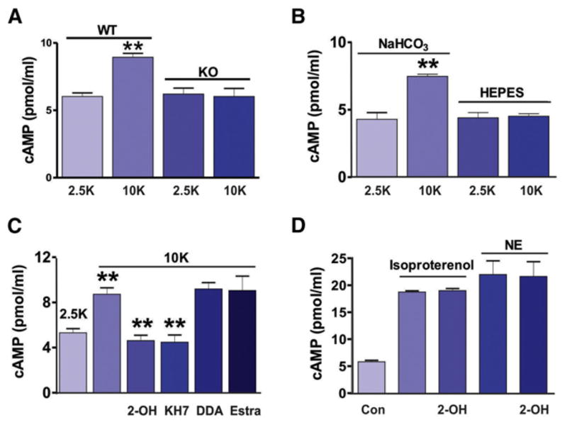
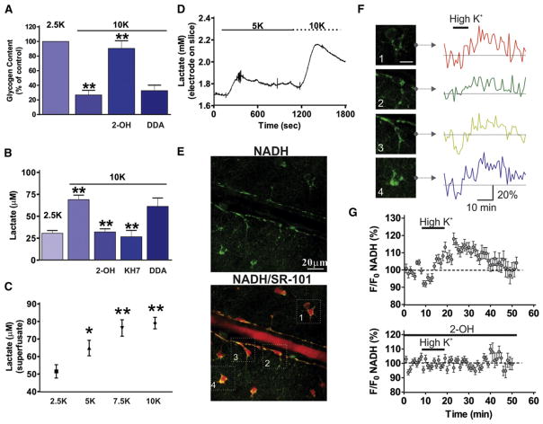
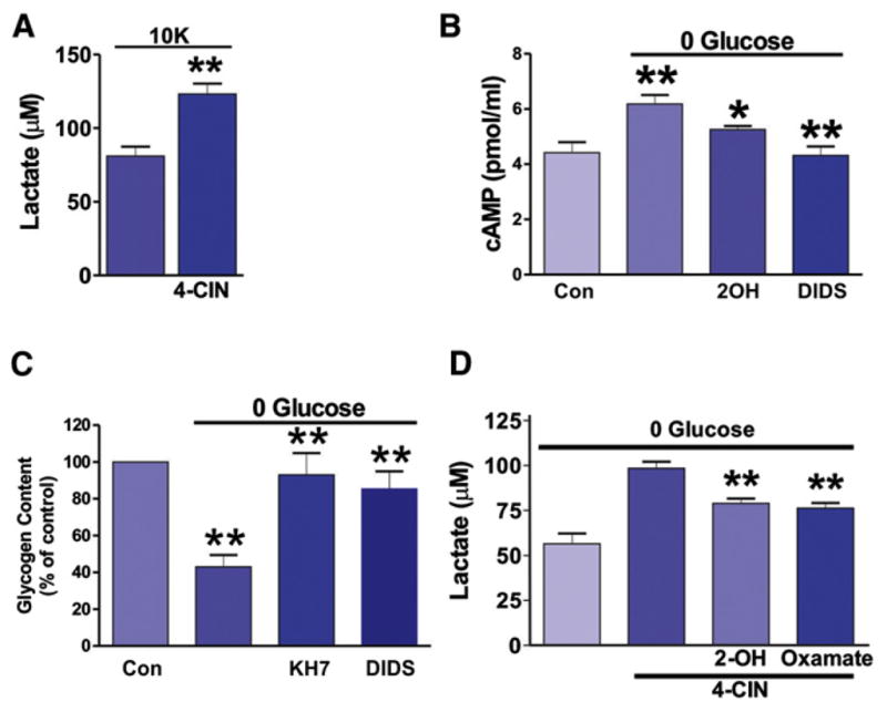
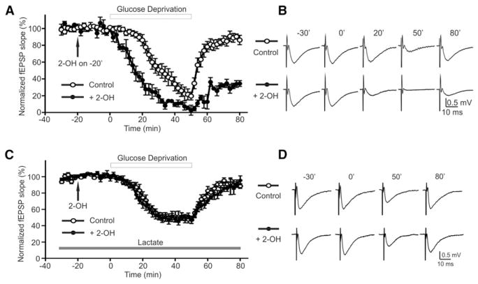
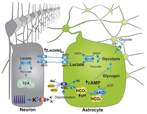
Astrocytes are proposed to participate in brain energy metabolism by supplying substrates to neurons from their glycogen stores and from glycolysis. However, the molecules involved in metabolic sensing and the molecular pathways responsible for metabolic coupling between different cell types in the brain are not fully understood. Here we show that a recently cloned bicarbonate (HCO₃⁻) sensor, soluble adenylyl cyclase (sAC), is highly expressed in astrocytes and becomes activated in response to HCO₃⁻ entry via the electrogenic NaHCO₃ cotransporter (NBC). Activated sAC increases intracellular cAMP levels, causing glycogen breakdown, enhanced glycolysis, and the release of lactate into the extracellular space, which is subsequently taken up by neurons for use as an energy substrate. This process is recruited over a broad physiological range of [K⁺](ext) and also during aglycemic episodes, helping to maintain synaptic function. These data reveal a molecular pathway in astrocytes that is responsible for brain metabolic coupling to neurons.


Figures

Figure 1
Figure 1. Expression of Bicarbonate-Responsive Soluble Adenylyl Cyclase in Astrocytes In Situ and In Vitro
(A and B) Immunohistochemical staining shows that GFAP-labeled astrocyte somata and major processes, including endfeet, expressed sAC (using R21, anti-sAC monoclonal antibody) (A), whereas MAP-2-labeled neuronal somata and dendrites revealed no specific sAC staining (B). (C) Western blots using sAC antibody (R21) show that sAC protein is found in both rat brain slices and cultured astrocytes. Blocking peptide disrupts antigen-antibody interaction between R21 and sAC. (D) RT-PCR results showing sAC mRNA is expressed in rat brain slices and cultured astrocytes. (E) sAC-immunoreactivity is found in glial processes in the stratum radiatum of the hippocampal CA1 region. Representative electron micrographs from a male wild-type mouse (left) demonstrate sAC-immunoperoxidase labeling in astrocytes (yellow outline) identified by glial filaments (gf) and other glial processes (GP), but not in a male Sacytm1Lex/Sacytm1Lex mouse (right; yellow highlight, unlabeled GP). S, synapse. (F) Quantification of the number of glial profiles (n = 3 mice per condition, ***p < 0.001). Error bars indicate SEM.
Figure 2
Figure 2. Activation of sAC Increases cAMP Concentration in Astrocytes In Vitro Detected by a FRET Sensor
(A) Pseudocolored cultured astrocytes expressing a cAMP FRET sensor. High [K+]ext increased the cAMP concentration (increased FRET ratio), which was blocked by the sAC-selective inhibitor 2-OH (20 μM). (B) Elevated [K+]ext (to 5 or 10 mM) increased FRET ratios over time, indicating increased intracellular cAMP levels, which were blocked by 2-OH or the NBC inhibitor DIDS (450 μM). (C) Summary data showing the increase of the FRET ratio either by changing the external solution from HCO3-free (with HEPES buffered) to one containing HCO3 (with regular aCSF solution) or by adding forskolin (25 μM) to stimulate tmACs. Error bars indicate SEM.
Figure 3
Figure 3. Activation of sAC Increases cAMP Concentration In Situ
(A) Raising [K+]ext to 10 mM significantly increased the cAMP level in brain slices from the wild-type mice but had no effect in brain slices from sAC KO mice. (B) ELISA showed high [K+]ext increased [cAMP] in rat brain slices only in the presence of HCO3. (C) ELISA demonstrating the increase of cAMP in high [K+]ext was reduced by sAC inhibitors, 2-OH (20 μM), or KH7 (10 μM) but not by the tmAC inhibitor DDA (50 μM). An inert estrogen parent compound, 17β-estradiol (a negative control for 2-OH), had no effect on the high K+-induced increase in cAMP. (D) ELISA showed 2-OH has no effect on cAMP production by the activation of beta-adrenoceptors using isoproterenol (100 μM) or norepinephrine (NE, 10 μM).
Figure 4
Figure 4. High [K+]ext Induces Glycogen Breakdown and Increased Lactate Production via sAC Activation
(A) High [K+]ext stimulated glycogen breakdown in brain slices, which was inhibited by the sAC inhibitor 2-OH (20 μM) but not by the tmAC inhibitor DDA (50 μM). (B) High [K+]ext increased lactate release from brain slices, which was blocked by KH7 (10 μM) and 2-OH but not DDA. (C) Lactate release from slices in response to different [K+]ext showed dose dependency. (D) Direct measurements using a lactate enzyme-based electrode showed the rapid time course of lactate release from brain slices. A transient increase of lactate was induced by 5 mM [K+]ext and the addition of 10 mM [K+]ext led to a further augmentation. High [K+]ext increased the cytosolic astrocyte NADH signal. (E) Hippocampal arteriole and astrocytes showing NADH (top) and colocalization with the astrocyte marker SR-101 (bottom). (F) NADH fluorescence changes in response to high [K+]ext in four astrocytes (from E). (G) Summary of NADH fluorescence changes in response to high [K+]ext (top) and block with 2-OH (bottom). Application of 10 mM [K+]ext increased the NADH signal (top), which was reduced by sAC inhibition with 2-OH (bottom).
Figure 5
Figure 5. sAC-Dependent Glycogen Breakdown and Lactate Delivery from Astrocytes to Neurons during Hypoglycemia
(A) Inhibition of neuronal uptake of lactate with 4-CIN increased extracellular lactate from brain slices in high [K+]ext. (B) Removing glucose from the aCSF for 15 min increased cAMP levels, which were inhibited by 2-OH (20 μM) and DIDS (450 μM). (C) Depletion of extracellular glucose significantly decreased glycogen content in rat brain slices and this was significantly inhibited by KH7 and DIDS. (D) 4-CIN in the absence of glucose increased extracellular lactate compared to glucose deprivation alone, which was inhibited by 2-OH (20 μM) and oxamate (2.5 mM).
Figure 6
Figure 6. sAC Activation during Hypogly-cemia Protects Synaptic Function by Lactate Release
(A) Glucose deprivation (open circle) decreased fEPSP amplitude, which recovered when glucose was reintroduced. fEPSP amplitude declined more rapidly to a greater extent and did not recover as well when sAC was inhibited with 2-OH (filled circle). (B) fEPSP traces from corresponding time points in (A). (C) Glucose deprivation (open circle) in the presence of exogenous lactate (5 mM). The adverse effect of 2-OH (filled circle) on fEPSP decline and recovery in hypoglycemia was no longer observed in the presence of lactate (5 mM). (D) fEPSP traces from corresponding time points in (C). Error bars indicate SEM.
Figure 7
Figure 7. A Summary Diagram Showing High [K+]ext-Mediated Activation of sAC via HCO3 Influx through NBCs and Its Physiological Role
A diagram showing high [K+]ext-mediated activation of sAC via HCO3 influx through NBCs and its role in increasing intracellular cAMP concentration, initiating glycogenolysis and producing lactate, which can be used as a neuronal fuel source.

References

    1. Alle H, Roth A, Geiger JR. Energy-efficient action potentials in hippocampal mossy fibers. Science. 2009;325:1405–1408. - PubMed
    1. Attwell D, Laughlin SB. An energy budget for signaling in the grey matter of the brain. J Cereb Blood Flow Metab. 2001;21:1133–1145. - PubMed
    1. Aubert A, Costalat R, Magistretti PJ, Pellerin L. Brain lactate kinetics: Modeling evidence for neuronal lactate uptake upon activation. Proc Natl Acad Sci USA. 2005;102:16448–16453. - PMC - PubMed
    1. Beierlein M, Gee KR, Martin VV, Regehr WG. Presynaptic calcium measurements at physiological temperatures using a new class of dextran-conjugated indicators. J Neurophysiol. 2004;92:591–599. - PubMed
    1. Bengtsson F, Boris-Möller F, Hansen AJ, Siesjö BK. Extracellular pH in the rat brain during hypoglycemic coma and recovery. J Cereb Blood Flow Metab. 1990;10:262–269. - PubMed

Publication types

MeSH terms