. 2012 Nov 25;433(2):506-12.
doi: 10.1016/j.virol.2012.08.044. Epub 2012 Sep 20.

Orthopoxvirus species and strain differences in cell entry

Affiliations

Orthopoxvirus species and strain differences in cell entry

Zain Bengali et al. Virology. .

Abstract

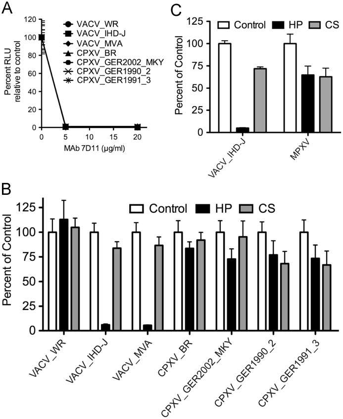
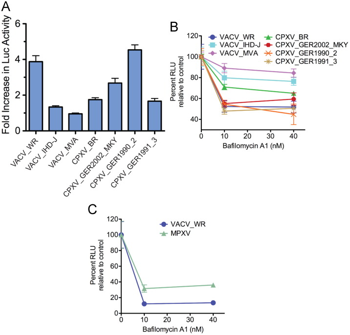
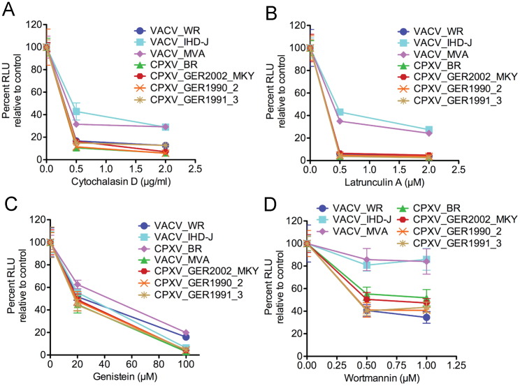
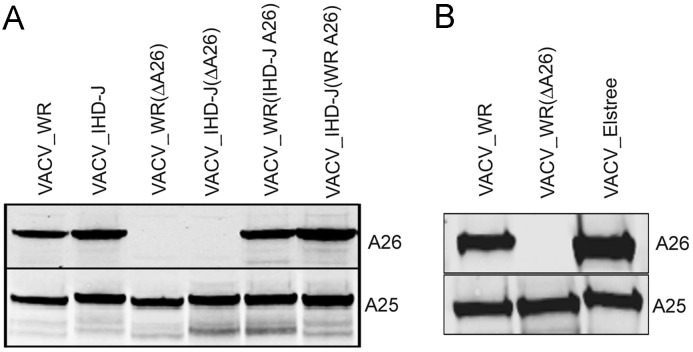
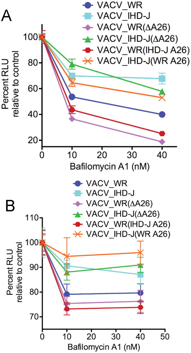
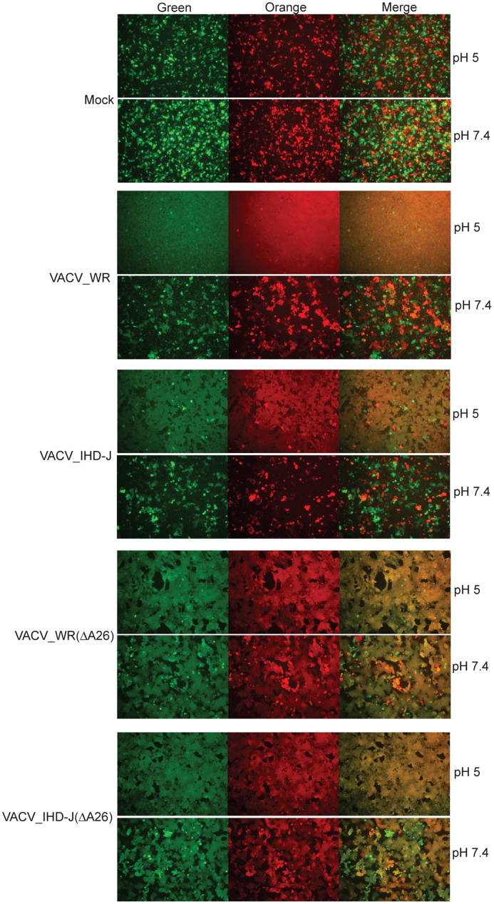
Vaccinia virus (VACV) enters cells by a low pH endosomal route or by direct fusion with the plasma membrane. We previously found differences in entry properties of several VACV strains: entry of WR was enhanced by low pH, reduced by bafilomycin A1 and relatively unaffected by heparin, whereas entry of IHD-J, Copenhagen and Elstree were oppositely affected. Since binding and entry modes may have been selected by specific conditions of in vitro propagation, we now examined the properties of three distinct, recently isolated cowpox viruses and a monkeypox virus as well as additional VACV and cowpox virus strains. The recent isolates were more similar to WR than to other VACV strains, underscoring the biological importance of endosomal entry by orthopoxviruses. Sequence comparisons, gene deletions and gene swapping experiments indicated that viral determinants, other than or in addition to the A26 and A25 "fusion-suppressor" proteins, impact entry properties.


Figures

Fig. 1
Fig. 1
Effects of neutralizing MAb, heparin and chondroitin sulfate on entry of recombinant LUC-expressing MVs. Purified MVs were incubated on ice with (A) 0, 5 or 20 μg/ml of MAb 7D11 for 30 min or (B and C) 0 or 50 μg/ml of heparin (HP) or chondroitin sulfate (CS) for 30 min. The treated MVs were adsorbed to BS-C-1 cells at 4 °C for 1 h in the presence of inhibitors and then incubated at 37 °C for 1 h. Cells were lysed and LUC activity determined. (C) MPXV experiments were conducted as described in the above panels except in a BSL-3 laboratory. Abbreviation: RLU, relative light units.
Fig. 2
Fig. 2
Effects of low pH and bafilomycin A1 treatment on cell entry of LUC-expressing recombinant MVs. (A) BS-C-1 cells were incubated with MVs at a multiplicity of 1 plaque forming unit (PFU) per cell at 4 °C for 1 h, followed by washing to remove unbound virus and exposure to pH 5 or pH 7.4 buffer for 3 min at 37oC. Cells were then washed and incubated at 37 °C at neutral pH for 1 h. Cells were lysed and LUC activity measured. The ratios of LUC activity following low and neutral pH are plotted for each virus. Error bars are shown. (B and C) BS-C-1 cells were pretreated with 0, 10 or 40 nM bafilomycin A1 for 1 h at 37 °C. Pretreated cells were incubated with indicated recombinant MVs in the presence of bafilomycin A1 for 1 h at 4 °C, unattached virus was removed by washing, and the cells were incubated at 37 °C at for 1 h in the absence or presence of bafilomycin A1. The cells were lysed and LUC activity measured. Abbreviation: RLU, relative light units.
Fig. 3
Fig. 3
Effects of cytochalasin D, latrunculin A, genestein and wortmannin on entry of LUC-expressing recombinant MVs. BS-C-1 cells were pretreated with (A) 0, 0.5 or 2.0 μg/ml of cytochalasin D; (B) 0, 0.5 or 2 μM latrunculin; (C) 0, 20 or 100 μM genestein; (D) 0, 0.5 or 1.0 μM wortmannin. Cells were infected and incubated as in Fig. 2 and LUC activity determined. The percent activity relative to 0 drug was plotted with error bars.
Fig. 4
Fig. 4
Detection of A26 and A25 in purified recombinant MVs. (A) Purified Luc-recombinant MVs of VACV strains WR and IHD-J, A26 deletion mutants VACV WR(ΔA26) and VACV IHD-J(ΔA26), and swap mutants VACV WR(IHD-J A26) and VACV IHD-J(WR A26) were analyzed by Western blotting with antibody to the VACV A26 protein and the CPXV ATI protein, which recognizes VACV A25 because of high sequence conservation. (B) VACV WR, VACV WR(ΔA26) and VACV Elstree MVs were analyzed as in panel A.
Fig. 5
Fig. 5
Effect of bafilomycin A1 on entry of LUC-expressing mutant viruses. BS-C-1 (A) and HeLa (B) cells were treated with bafilomycin A1, infected with LUC-expressing WR and IHD-J or A26 deletion mutants or swap mutants defined in the legend to Fig. 4. LUC activity was measured as described in the legend to Fig. 2.
Fig. 6
Fig. 6
Fusion from without. HeLa cells were separately transfected with plasmid expressing CMV-promoter controlled orange fluorescent protein or green fluorescent protein. After 24 h, the cells were harvested, mixed in 1:1 ratio and plated together in a 96-well plate. After forming a monolayer, the cells were infected with 100 PFU of purified virus per cell, treated with either pH 7.4 or 5.0 buffer for 2 min, and incubated in neutral pH medium for additional 1 h at 37 °C. Fluorescent cells were detected with a Leica DM IRB fluorescent microscope.
None

Similar articles

Cited by

References

    1. Armstrong J.A., Metz D.H., Young M.R. The mode of entry of vaccinia virus into L cells. J. Gen. Virol. 1973;21:533–537. - PubMed
    1. Bengali Z., Townsley A.C., Moss B. Vaccinia virus strain differences in cell attachment and entry. Virology. 2009;389:132–140. - PMC - PubMed
    1. Bisht H., Weisberg A.S., Moss B. Vaccinia virus L1 protein is required for cell entry and membrane fusion. J. Virol. 2008;82:8687–8694. - PMC - PubMed
    1. Breman J.G., Kalisa R., Steniowski M.V., Zanotto E., Gromyko A.I., Arita I. Human monkeypox, 1970–79. Bull. WHO. 1980;58:165–182. - PMC - PubMed
    1. Brown E., Senkevich T.G., Moss B. Vaccinia virus F9 virion membrane protein is required for entry but not virus assembly, in contrast to the related l1 protein. J. Virol. 2006;80:9455–9464. - PMC - PubMed

Publication types

MeSH terms