

doi: 10.1016/j.ajog.2012.08.036.
Epub 2012 Sep 7.
See it in 3D!: researchers examined structural links between the cardinal and uterosacral ligaments
Affiliations
- PMID: 22999154
- PMCID: PMC3515066
- DOI: 10.1016/j.ajog.2012.08.036
Item in Clipboard
See it in 3D!: researchers examined structural links between the cardinal and uterosacral ligaments
Am J Obstet Gynecol.
2012 Nov.
No abstract available
Conflict of interest statement
All other authors report no conflict of interest.
Figures
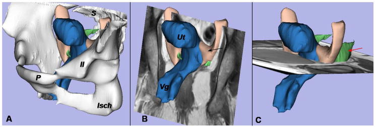
A 3- dimensional (3D) model of the cardinal ligament and uterosacral ligament is oriented relative to the coronal and axial planes. A, The bony pelvis, including the pubis (P), ilium (Il), ischium (Isch), and sacrum (S), is displayed. B, Positioning of the uterus (Ut), cardinal ligament (black arrow), and vagina (Vg) can be seen in this coronal image. C, Note the location of the uterosacral ligament (red arrow) in this axial MR scan.
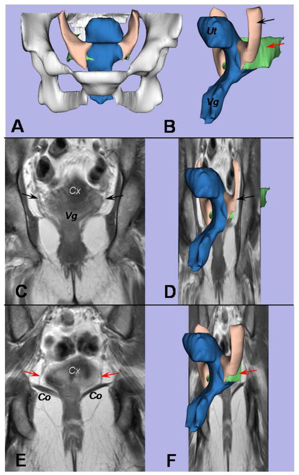
A coronal view of the cardinal and uterosacral ligaments is provided. A, The bony pelvis can be seen in this front view of the 3D model. B, An oblique left-side view displays the uterus (Ut) and vagina (Vg) in blue, the uterosacral ligament in green (red arrow), and the cardinal ligament in cream (black arrow). C, A coronal scan is at the level of the cardinal ligament; the cervix (Cx) and vagina (Vg) are marked. D, This is where the structures in the 3D model shown in Figure 2B would be situated in the plane. E, The uterosacral ligament (red arrows) can be seen when the view is moved 10 mm dorsal from where the cardinal ligament is seen in Figure 2C. The cervix (Cx) and coccygeus muscle (Co) are shown. F, Again, the 3D model shown in Figure 2B is shown within the coronal plane.
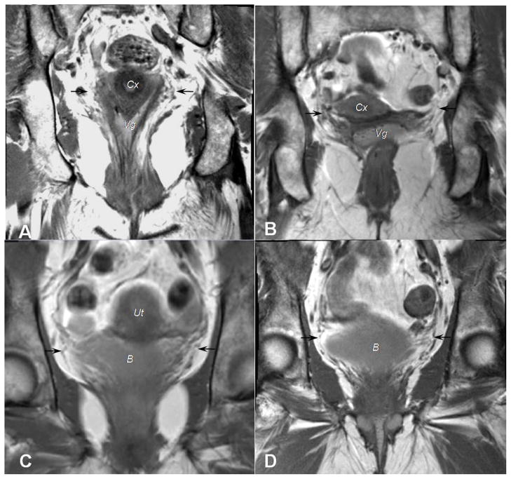
Coronal scans from 4 different women illustrate attachments of the cardinal ligament to the pelvic organs. A, B, Black arrows point to the cardinal ligament’s insertions at both the cervix (Cx) and upper vagina (Vg). C,D, Fibers of the cardinal ligament, again identified with black arrows, continue to the bladder (B).
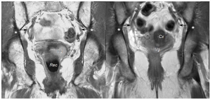
On coronal scans of 2 different subjects, you can see the region of the cardinal ligament’s attachment to the pelvic wall (black arrow) near the origin of the internal iliac artery’s anterior trunk. This was seen in 7 patients. For 13 patients, the region of attachment was located at the upper level of the great sciatic foramen, as identified with asterisks. The vagina (Vg), rectum (Rec), and cervix (Cx) are labeled.
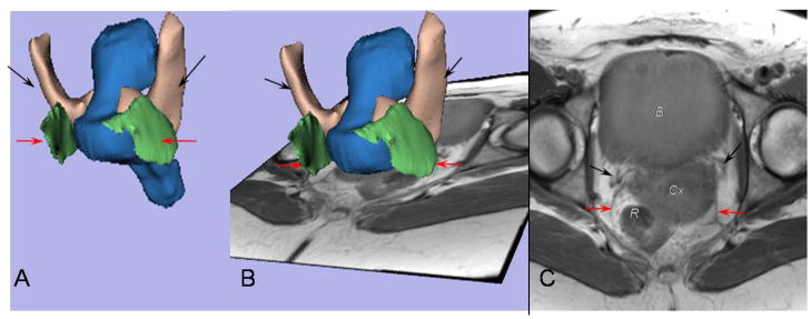
The cardinal and uterosacral ligaments mingle near the cervix and upper vagina. A, This is the back of the 3D model. The red arrows identify the uterosacral ligament, and the black arrows show the cardinal ligament. B, This MR scan is at the level of the cervix. C, Fibers of both ligaments converge towards the cervical-vaginal junction, where they are in close continuity. The bladder (B), the cervix (Cx), and the rectum (R) are noted.
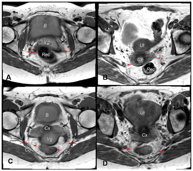
Attachments of the uterosacral ligament on an axial MR image are viewed in scans from 4 different subjects. A, Ventral attachments are displayed. The black arrows indicate the cardinal ligament. Deep uterosacral fibers, as denoted by the red arrows, are inserted at the upper vagina. B, Again, ventral attachments are shown. The black arrows point to the cardinal ligament. Superficial uterosacral fibers, at the red arrowheads, are inserted at the cervix. C, Dorsal attachments are shown. The sacrospinous ligament-coccygeus muscle insertion of the deep uterosacral fibers are designated by red arrows; superficial uterosacral fibers, the red arrowheads. D, Another view of dorsal attachments is displayed. Presacral insertion of the uterosacral ligament is identified with red arrows. The bladder (B), vagina (Vg), rectum (Rec), cervix (Cx), uterus (Ut), sacrum (S), and coccygeus muscle (Co) are identified in different views.
References
-
- Rooney K, Kenton K, Mueller ER, FitzGerald MP, Brubaker L. Advanced anterior vaginal wall prolapse is highly correlated with apical prolapse. Am J Obstet Gynecol. 2006;195:1837–40. - PubMed
-
- DeLancey JO. Anatomic aspects of vaginal eversion after hysterectomy. Am J Obstet Gynecol. 1992;166(6Pt1):1717–24. - PubMed
-
- Campbell RM. The anatomy and histology of the sacrouterine ligaments. Am J Obstet Gynecol. 1950;59:1–12. - PubMed
-
- Range RL, Woodburne RT. The gross and microscopic anatomy of the transverse cervical ligament. Am J Obstet Gynecol. 1964;90:460–7. - PubMed
Publication types
MeSH terms
Grants and funding
LinkOut - more resources
Full Text Sources
Medical
