. 2012 Oct 18;12(4):509-20.
doi: 10.1016/j.chom.2012.08.004. Epub 2012 Sep 20.

Outer membrane vesicles of a human commensal mediate immune regulation and disease protection

Affiliations

Outer membrane vesicles of a human commensal mediate immune regulation and disease protection

Yue Shen et al. Cell Host Microbe. .

Abstract

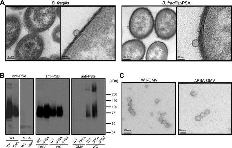
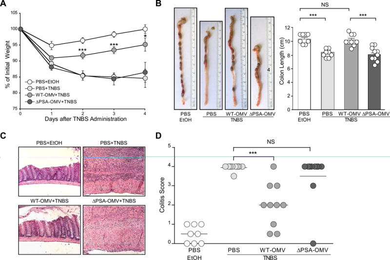
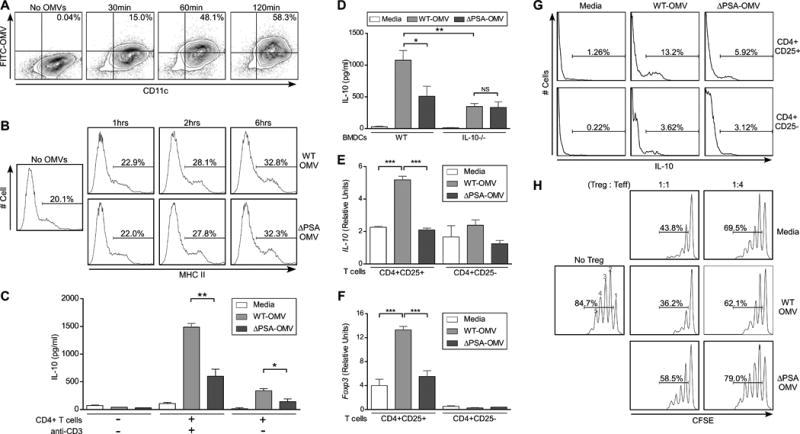
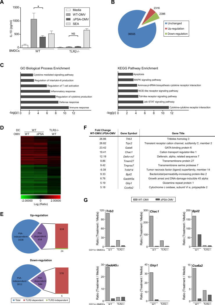
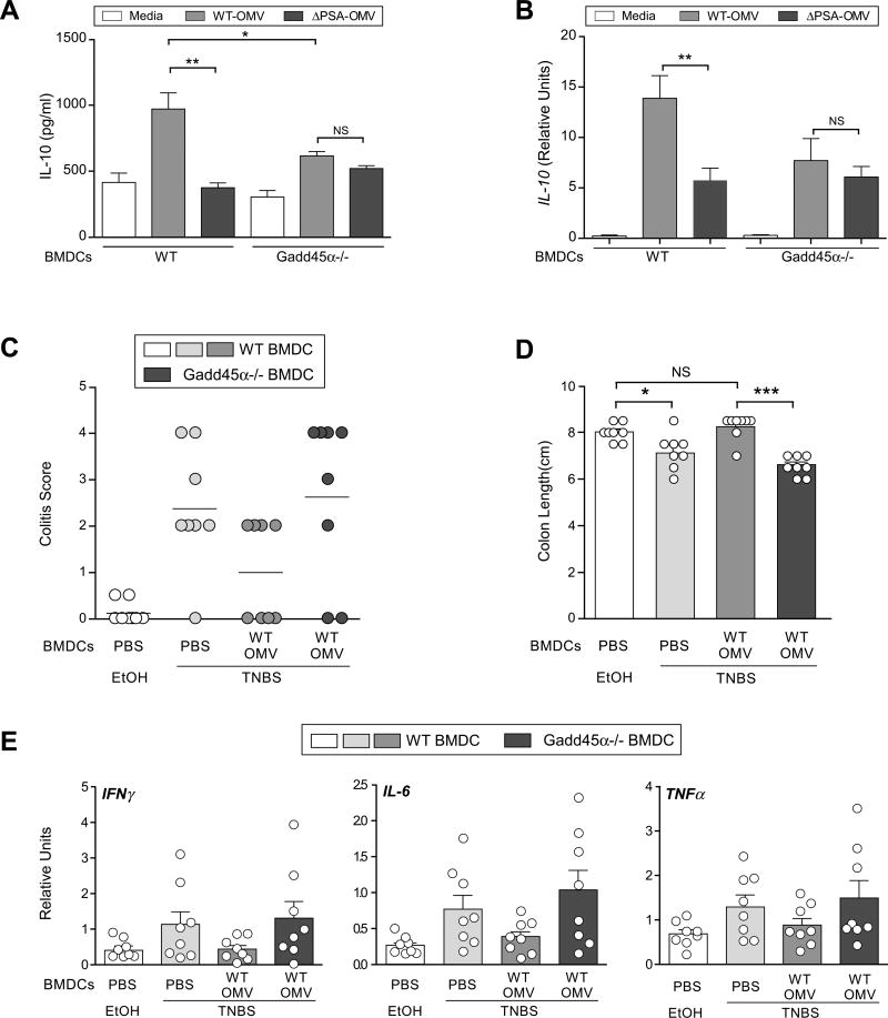
Commensal bacteria impact host health and immunity through various mechanisms, including the production of immunomodulatory molecules. Bacteroides fragilis produces a capsular polysaccharide (PSA), which induces regulatory T cells and mucosal tolerance. However, unlike pathogens, which employ secretion systems, the mechanisms by which commensal bacteria deliver molecules to the host remain unknown. We reveal that Bacteroides fragilis releases PSA in outer membrane vesicles (OMVs) that induce immunomodulatory effects and prevent experimental colitis. Dendritic cells (DCs) sense OMV-associated PSA through TLR2, resulting in enhanced regulatory T cells and anti-inflammatory cytokine production. OMV-induced signaling in DCs requires growth arrest and DNA-damage-inducible protein (Gadd45α). DCs treated with PSA-containing OMVs prevent experimental colitis, whereas Gadd45α(-/-) DCs are unable to promote regulatory T cell responses or suppress proinflammatory cytokine production and host pathology. These findings demonstrate that OMV-mediated delivery of a commensal molecule prevents disease, uncovering a mechanism of interkingdom communication between the microbiota and mammals.


Figures

Figure 1
Figure 1
Outer membrane vesicles (OMVs) from Bacteroides fragilis contain PSA. (A) Transmission electron microscopy of electron dense layer (EDL) enriched B. fragilis (WT-OMV) and B. fragilisΔPSA (ΔPSA-OMV) reveals vesicles budding from the bacterial surface. (B) Immunoblot analysis of whole cell (WC) and OMV extracts ofbacteria show that PSA and PSB are associated with vesicles, while PSG is only found associated with the bacterial cell. Deletion mutants for PSA (ΔPSA), PSB (ΔPSB) and PSG (ΔPSG) confirm specificity of each anti-sera. (C) Immunogold labeling of purified OMVs, stained with anti-PSA and anti-IgG-colloidal gold conjugate (5nm), analyzed by electron microscopy shows specific staining for PSA only in OMVs from wild-type bacteria but not B. fragilisΔPSA. See also Figure S1.
Figure 2
Figure 2
OMVs protect animals from experimental colitis and intestinal inflammation in a PSA-dependent manner. (A) Weight loss in animals following the induction of TNBS colitis (day 0) measured as reduction from initial weight until day of sacrifice (day 4). All groups contained at least 4 animals, with error bars indicating standard error (SEM). Results are representative of 3 independent trials. * p<0.05; *** p<0.001. (B) Images of colons immediately following resection and quantification of length (indicated in the bar graph) from vehicle treated (EtOH) and TNBS groups (n=4 animals/group). Error bars indicate SEM. Results are shown from 3 combined experiments where each was performed independently. *** p<0.001. NS: not significant. (C) Images from hematoxylin and eosin (H & E) stained colon sections representative of each treatment group. (D) Colitis scores from animals assigned by a blinded pathologist (G.W.L) according to a standard scoring system (see Experimental Procedures). Each symbol represents an individual animal. Results are shown from 3 combined experiments, each performed independently. *** p<0.001. NS: not significant. See also Figure S2.
Figure 3
Figure 3
Treatment of DCs with PSA-containing OMVs induces IL-10 production and Foxp3 expression from T cells. (A) Flow cytometry (FC) analysis of OMV internalization by DCs. OMVs were labeled with FITC (Fluorescein isothiocyanate) and incubated with cultured DCs for various times. Cells were stained with anti-CD11c. Percentages show CD11c+OMV+ cell populations. (B) FC plots of DCs incubated with WT-OMVs and ΔPSA-OMVs for various times and stained with anti-CD11c and anti-MHCII (Major histocompatibility complex II). Percentages show MHCII+ populations among CD11c+ cells. (C) ELISA analysis for IL-10 production from culture supernatants of DCs or DC-T cell co-cultures, where DCs were pulsed with OMVs for 18 hours, washed and incubated with or without splenic CD4+ T cells. Supernatants were collected at day 4 of culture. Media samples indicate DCs that were not pulsed with OMVs, but otherwise treated identically. Anti-CD3 was added to some samples to demonstrate T cell-specific responses. Error bars indicate SEM from quadruplicate samples. Results are representative of over 5 independent trials. * p<0.05; ** p<0.01. (D) ELISA analysis of DC-T cell co-cultures similar to (C), with IL-10-/- DCs. Error bars indicate SEM from quadruplicate samples. Results are representative of 3 independent trials. * p<0.05. ** p<0.01. NS: not significant. (E, F) Transcript levels of IL-10 (E) and Foxp3 (F) of RNA recovered from purified CD4+ T cell subsets following in vitro culture with DCs. Co-cultures were set up as in (C); on day 4, CD4+CD25+ and CD4+CD25- T cells were purified by magnetic bead separation (>95% purity) and RNA extracted for qRT-PCR. Relative values were normalized to β-actin. Error bars indicate SEM. Results are shown from 3 combined experiments each performed independently. * p<0.05; *** p<0.001. NS: not significant. (G) FC histograms of IL-10 expression by CD4+ T cell subsets following 4 days of co-culture with DCs treated with OMVs. Splenic CD4+ T cell were purified from IL-10-GFP mice, stained with anti-CD4 and anti-CD25 following co-culture, and IL-10 expression measured by GFP expression. Percentages show IL-10+ populations amongCD4+CD25+ and CD4+CD25- subsets. Results are representative of 2 independent trials. (H)In vitro suppression of naïve responder cells by purified CD4+CD25+ T cells following co-culture with DCs treated with media (control), WT-OMVs and ΔPSA-OMVs. CD4+CD25-responder cells (effector cells; Teff) were pulsed with CFSE, incubated with TREGS and stimulated with anti-CD3 and APC (CD4+ T cell depleted splenocytes) for 3 days. Cell proliferation was measured by FC as a function of CFSE dilution. TREG:Teff ratios are indicated, and percentages show total proliferating cells. No TREG: CD4+CD25- cells only. Numbers above peaks represent number of cell divisions. Results are representative of 3 independent trials. See also Figure S3.
Figure 4
Figure 4
TLR2 on DCs is required to sense OMV-associated PSA and induces genes specific for OMVs. (A) ELISA analysis of DC-T cell co-cultures similar to Figure 3(C) from WT and TLR2-/- DCs. SEA: staphylococcal enterotoxin A. Error bars indicate SEM from quadruplicate samples. Results are representative of 3 independent trials. * p<0.05. NS: not significant. (B) Pie chart showing the number of up-regulated genes (red) and down-regulated genes (green) upon stimulation with PSA-containing OMVs during in vitro cultured with CD11c+ BMDCs, using a Whole Mouse Genome Microarray analysis. Unaffected genes (blue). Only genes with a p value <0.01 and fold change >2 were used for subsequent analysis. (C) Gene ontology analysis of changes in gene expression levels in BMDCs for various biological processes or pathways. (D) Heat-map analysis of gene expression in BMDCs from either wild-type or TLR2-/- animals upon OMV stimulation reveals that approximately 30% of the genes are either up-regulated or down-regulated in a PSA dependent manner and that the majority of the genes are TLR2 dependent. (E) Pie charts show that the majority of the genes in BMDCs that respond to PSA are dependent on TLR2. (F) List of select genes of highest fold change of expression level upon OMV stimulation in a PSA- and TLR2-dependent manner. (G) qRT-PCR analysis of the expression level of genes listed in (F) identified and confirmed 6 genes that are significantly up-regulated by OMVs in a PSA- and TLR2-dependent manner from purified CVD11c+ BMDCs. Results are representative of 3 independent experiments. See also Figure S4.
Figure 5
Figure 5
Gadd45α on DCs is required for in vitro IL-10 production and protection from colitis by OMVs. (A) ELISA analysis of DC-T co-cultures similar to Figure 3(C), but also including Gadd45α-/- DCs. Error bars indicate SEM from quadruplicate samples. Results are representative of 3 independent trials. *p<0.05. ** p<0.01. NS: not significant. (B) qRT-PCR analysis of IL-10 transcript levels from either WT or Gadd45α-/- BMDCs after 16-18hrs incubation with OMVs in vitro. Error bars indicate SEM from triplicate samples. Results are representative of 3 independent trials. ** p<0.01. NS: not significant. (C) Colitis scores from DC recipient animals induced for TNBS colitis. Each symbol represents an individual animal. Results are shown from 2 combined experiments, each performed independently. (D) Colon length from mice, at day 2 following TNBS treatment. Each symbol represents an individual animal. Error bars indicate SEM from 8 animals. Results are shown from 2 combined experiments each performed independently. * p<0.05. *** p<0.001. NS: not significant. (E) Cytokine analysis by qRT-PCR of RNA recovered from whole colon. Each symbol represents an individual animal. Error bars indicate SEM from 8 animals. Results are shown from 2 combined experiments, each performed independently. See also Figure S5.

Comment in

References

    1. Beveridge TJ. Structures of gram-negative cell walls and their derived membrane vesicles. J Bacteriol. 1999;181:4725–4733. - PMC - PubMed
    1. Buckner JH. Mechanisms of impaired regulation by CD4(+)CD25(+)FOXP3(+) regulatory T cells in human autoimmune diseases. Nature reviews Immunology. 2010;10:849–859. - PMC - PubMed
    1. Cerdeno-Tarraga AM, Patrick S, Crossman LC, Blakely G, Abratt V, Lennard N, Poxton I, Duerden B, Harris B, Quail MA, et al. Extensive DNA inversions in the B. fragilis genome control variable gene expression. Science. 2005;307:1463–1465. - PubMed
    1. Chung H, Pamp SJ, Hill JA, Surana NK, Edelman SM, Troy EB, Reading NC, Villablanca EJ, Wang S, Mora JR, et al. Gut immune maturation depends on colonization with a host-specific microbiota. Cell. 2012;149:1578–1593. - PMC - PubMed
    1. Coyne MJ, Tzianabos AO, Mallory BC, Carey VJ, Kasper DL, Comstock LE. Polysaccharide biosynthesis locus required for virulence of Bacteroides fragilis. Infect Immun. 2001;69:4342–4350. - PMC - PubMed

Publication types

MeSH terms

Associated data