Review
. 2012 Dec;22(6):733-42.
doi: 10.1016/j.sbi.2012.08.004. Epub 2012 Sep 19.

Crystal structures of 70S ribosomes bound to release factors RF1, RF2 and RF3

Affiliations
Review

Crystal structures of 70S ribosomes bound to release factors RF1, RF2 and RF3

Jie Zhou et al. Curr Opin Struct Biol. 2012 Dec.

Abstract

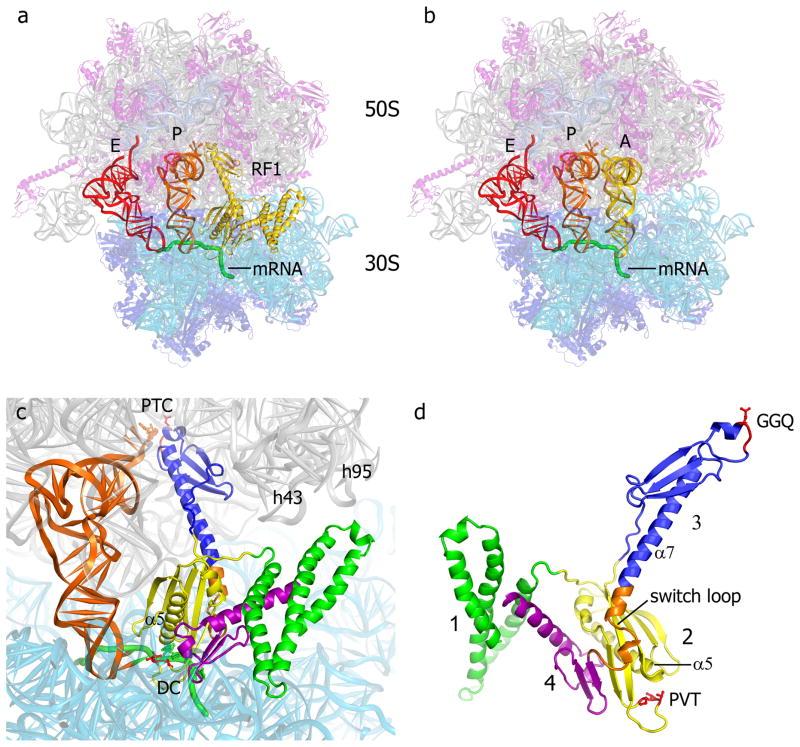
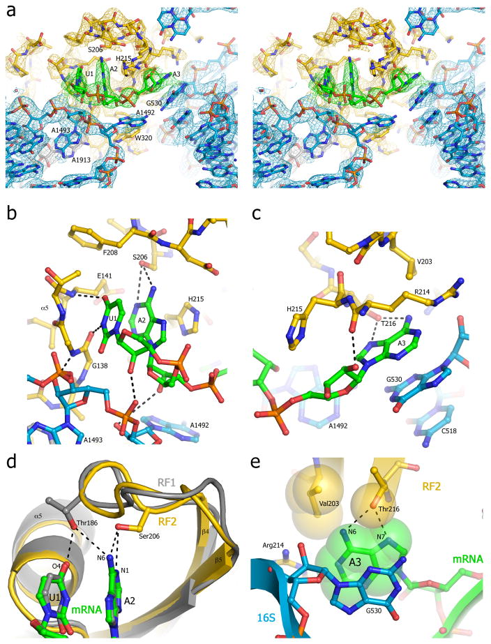
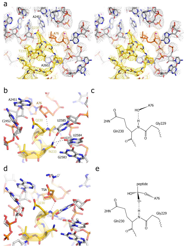
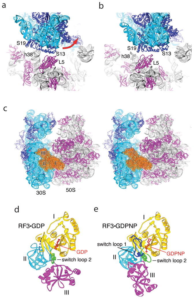
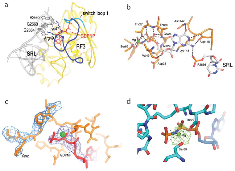
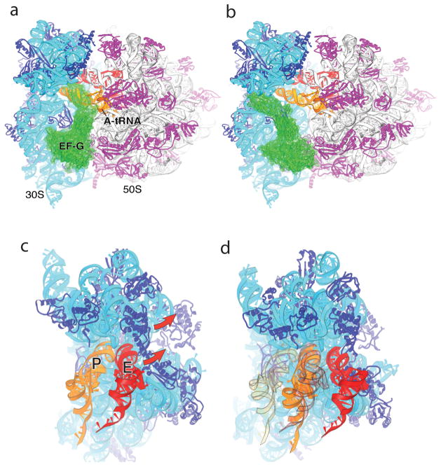
Termination is a crucial step in translation, most notably because premature termination can lead to toxic truncated polypeptides. Most interesting is the fact that stop codons are read by a completely different mechanism from that of sense codons. In recent years, rapid progress has been made in the structural biology of complexes of bacterial ribosomes bound to translation termination factors, much of which has been discussed in earlier reviews [1-5]. Here, we present a brief overview of the structures of bacterial translation termination complexes. The first part summarizes what has been learned from crystal structures of complexes containing the class I release factors RF1 and RF2. In the second part, we discuss the results and implications of two recent X-ray structures of complexes of ribosomes bound to the translational GTPase RF3. These structures have provided many insights and a number of surprises. While structures alone do not tell us how these complicated molecular assemblies work, is it nevertheless clear that it will not be possible to understand their mechanisms without detailed structural information.


Figures

Fig. 1
Fig. 1. Binding of RF1 to the 70S ribosome
(a) Positions of RF1 (yellow), P-site tRNA (orange), E-site tRNA (red) and a mRNA (green) bound to the 70S ribosome [23S rRNA (gray), 5S rRNA (light blue), 16S rRNA (cyan), 50S proteins (magenta) and 30S proteins (dark blue)]. (b) The position of A-site tRNA (yellow) in a 70S elongation complex [54] is similar to that of domains 2, 3 and 4 of RF1. (c) Orientation of RF1 in the A site of the 70S ribosome, along with the P-site tRNA (orange). The peptidyl transferase center (PTC), decoding center (DC) and helices h43 (the L11 stalk) and h95 (the sarcin-ricin loop) of 23S rRNA are indicated. (d) The structure of RF1 in its ribosome-bound conformation. The view is rotated approximately 180° from that shown in panels (a) and (c), for clarity. Domains 1, 2, 3 and 4 are shown in green, yellow, blue and magenta, respectively. The conserved GGQ and PVT motifs are shown in red, and the switch loop (see text) is in orange. From ref. [16].
Fig. 2
Fig. 2. Recognition of stop codons by RF1 and RF2
(a) Stereo view of the σA-weighted 3Fo-2Fc electron density map of the stop codon and surrounding elements of RF2 and the ribosome [17]. Electron density is contoured at 1.0 σ for RF2, and at 1.5 σ for rRNA and mRNA, and colored yellow (RF1), green (mRNA) and blue (16S rRNA). (b) Interaction of the hydroxyl group of Ser206 of the SPF motif of RF2 with A2 of the stop codon; (c) Interaction of Thr216 of RF2 with A3 of the stop codon; (d) Comparison of the positions of Thr186 of the PVT motif of RF1 (gray)[16] and Ser206 of the SPF motif of RF2 (yellow)[17], showing their respective modes of recognition of U1 and A2 of the UAA stop codon. (e) Packing of RF2 around A3 of the UAA stop codon. Val203 would be positioned to exclude water from H-bonding to O6 of guanine, helping to discriminate against guanine at position 3. The structure model is represented as Van der Waals surfaces in yellow (RF2), green (mRNA) and blue (16S rRNA). From ref. [17].
Fig. 3
Fig. 3. Interactions of the universally conserved GGQ motif in the peptidyl transferase center (PTC) of the termination complex
(a) Stereo view of σA-weighted 3Fo-2Fc electron density map for the PTC. Density for RF1 (yellow), P-tRNA (orange) and 23S rRNA (grey) was contoured at 1.7 σ. (b) Interaction of the backbone amide nitrogen of the universally conserved Gln230 with the 3′-OH of A76 of P-site tRNA. (c) Model for proposed product stabilization of the peptide release reaction by H-bonding between the main-chain amide nitrogen of Gln230 and the 3′-OH of A76. (d) Superposition of the structure of the peptidyl-transferase transition-state analog (TSA, orange) complexed with the 50S subunit (grey)[55] on the structure of the termination complex (this work). The main-chain amide nitrogen of Gln230 is positioned to form a hydrogen bond with the oxyanion of the TSA. (e) Model for potential transition-state stabilization of the peptide release reaction by H-bonding between RF1 and the TSA. From ref. [16].
Fig. 4
Fig. 4. Effects of binding RF3 to the 70S ribosome
(a,b) Large-scale rotation of the 30S subunit head induced by RF3 in the E. coli complex [43]. (a) In the classical-state ribosome [17], bridge B1a is formed between protein S13 and helix 38 of 23S rRNA, and B1b between S13 and L5. (b) In the RF3 complex, a large-scale (14°) counter-clockwise rotation of the 30S head results in disruption of bridges B1a and B1b; a new bridge R1b is formed between proteins S13 and S19 from the 30S subunit and L5 from the 50S subunit, involving a 34 Å displacement of the intersubunit contacts. (c) Stereo view of RF3 (orange) bound at the entrance to the interface cavity of the 70S ribosome. (d,e) Comparison of crystal structures of (d) free RF3·GDP [42] and (e) ribosome-bound RF3·GDPNP. A large-scale rotational movement of domain III (magenta) and smaller movements in domains I (yellow) and II (cyan) can be seen. Disordered segments in RF3·GDP are shown as dotted lines. Rearranged segments in RF3·GDPNP containing switch loops I and II are shown in blue and green, respectively. GDP and GDPNP are shown in red. From ref. [43].
Fig. 5
Fig. 5. Ribosome-induced ordering of the GTPase active site of RF3 in the E. coli complex
(a) Ordering of residues 39–69 of RF3 and re-positioning residues 70–73 (including the switch loop 1 residues 64–72) creates contacts between the G domain of RF3 and the SRL of 23S rRNA. Residues 39–63 are shown in dark blue, the switch loop in cyan, and the rest of RF3 in yellow. (b) An intricate network of H-bonds is formed between the base, ribose and phosphate groups of GDPNP and domain I of RF3, including elements of switch I (residues 64–72), P loop (residues 21–28), NKXD motif (residues142–145) and phosphate 2656 of the SRL of 23S rRNA. A Mg atom (green) is coordinated by Thr27, Ser91 and the β and γ phosphates of GDPNP (see Fig. S9). (c) His92 of RF3 is positioned at a distance of 8 Å from the γ phosphate of GDPNP. The 2Fo-Fc electron density map is shown around His92 and GDPNP, contoured at 1.3 σ and 2.3 σ, respectively. (d) Difference electron density showing Mg bound to GDPNP. An Fo-Fc difference map was calculated using the complete RF3·GDPNP·70S ribosome model, shown here contoured at 4.5 σ. The Mg ion (green) is coordinated with the β- and γ-phosphate oxygens of GDPNP and with the side-chain hydroxyls of Thr27 and Ser 69. From ref. [43].
Fig. 6
Fig. 6. Implications for EF-G and translocation
(a) Observed orientation of EF-G (green) bound to the classical-state 70S ribosome·EF-G complex [56], in which tRNA has been modeled into the A site; note the severe steric clash between domain IV of EF-G and A-site tRNA. (b) Orientation of EF-G in the classical-state ribosome, but with its domain I docked on the 50S subunit in the same orientation as observed for RF3·GDPNP in the E. coli structure [43]. Note the absence of steric clash with A-site tRNA or the ribosome. (c,d) P-site ASL contacts in the 30S subunit head are displaced by 23 Å in the RF3 complex, relative to their position in the classical-state complex. Docking of P-site (orange) and E-site (red) tRNAs on the (c) classical-state RF2 and (d) rotated RF3 structures illustrates that the combined 30S head and body rotations seen in the E. coli RF3 structure are sufficient to translocate an ASL from the 30S P site to the E site, while allowing passage of the ASL of P-site tRNA to the E site without clash with the ribosome. The views in (c) and (d) are shown in exactly the same orientation relative to the 50S subunit. In (d), the initial positions of the P- and E-site tRNAs as in (c) are shown in transparent orange and red, respectively. From ref. [43].

References

    1. Korostelev AA. Structural aspects of translation termination on the ribosome. RNA. 2011;17:1409–1421. - PMC - PubMed
    1. Klaholz BP. Molecular recognition and catalysis in translation termination complexes. Trends Biochem Sci. 2011;36:282–292. - PubMed
    1. Loh PG, Song H. Structural and mechanistic insights into translation termination. Curr Opin Struct Biol. 2010;20:98–103. - PubMed
    1. Dunkle JA, Cate JH. Ribosome structure and dynamics during translocation and termination. Annu Rev Biophys. 2011;39:227–244. - PubMed
    1. Petry S, Weixlbaumer A, Ramakrishnan V. The termination of translation. Curr Opin Struct Biol. 2008;18:70–77. - PubMed

Publication types

MeSH terms

Substances

LinkOut - more resources