. 2012 Sep 27;2(3):695-704.
doi: 10.1016/j.celrep.2012.08.015. Epub 2012 Sep 20.

High-throughput screening for bioactive molecules using primary cell culture of transgenic zebrafish embryos

Affiliations

High-throughput screening for bioactive molecules using primary cell culture of transgenic zebrafish embryos

Haigen Huang et al. Cell Rep. .

Abstract

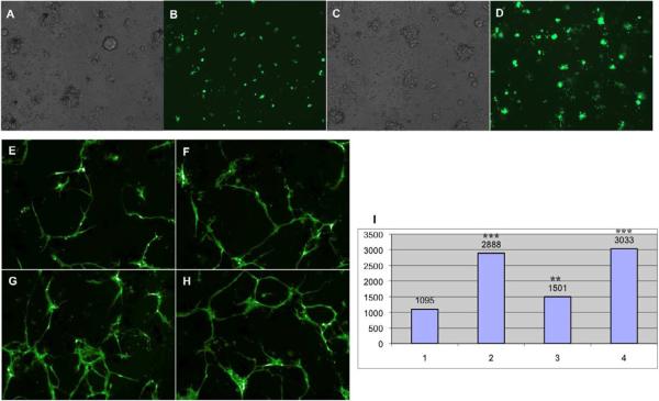
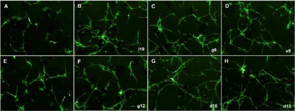
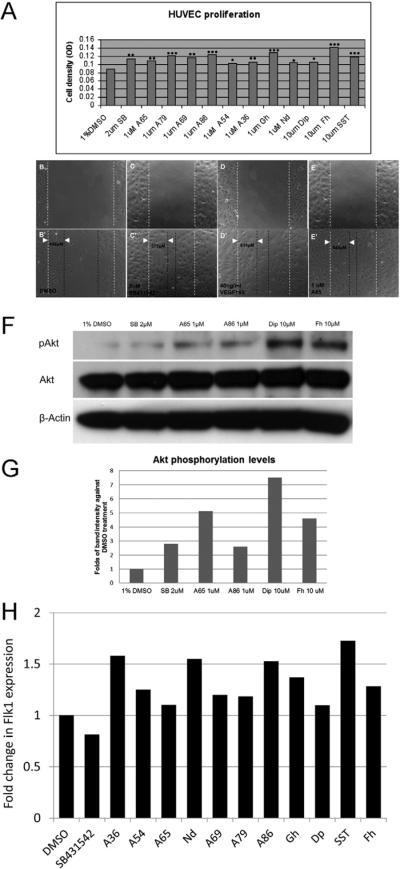
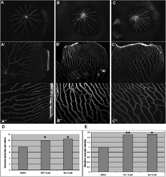
Transgenic zebrafish embryos expressing tissue-specific green fluorescent protein (GFP) can provide an unlimited supply of primary embryonic cells. Agents that promote the differentiation of these cells may be beneficial for therapeutics. We report a high-throughput approach for screening small molecules that regulate cell differentiation using lineage-specific GFP transgenic zebrafish embryonic cells. After validating several known regulators of the differentiation of endothelial and other cell types, we performed a screen for proangiogenic molecules using undifferentiated primary cells from flk1-GFP transgenic zebrafish embryos. Cells were grown in 384-well plates with 12,128 individual small molecules, and GFP expression was analyzed by means of an automated imaging system, which allowed us to screen thousands of compounds weekly. As a result, 23 molecules were confirmed to enhance angiogenesis, and 11 of them were validated to promote the proliferation of mammalian human umbilical vascular endothelial cells and induce Flk1+ cells from murine embryonic stem cells. We demonstrated the general applicability of this strategy by analyzing additional cell lineages using zebrafish expressing GFP in pancreatic, cardiac, and dopaminergic cells.


Figures

Figure 1
Figure 1
Overall strategy of primary cell-based high throughput screening for bioactive small molecules using cells dissociated from early transgenic zebrafish embryos.
Figure 2
Figure 2
Differentiation of flk1-GFP labeled endothelial cells in primary culture. Flk1-GFP expressing endothelial cells are scattered at day 1 and become flatten and elongated at day 2–4, undergoing tube formation. After 5–6 days of culture, GFP positive cells start to die (in blue circles). A and B show DIC and Hoechst 33342 (5 μg/ml for 4.5–5 hours) staining on primary cells at day 6.
Figure 3
Figure 3
Flk1-GFP labeled endothelial cells respond to growth factors and small molecules in primary culture. Endothelial cell proliferation and differentiation derived from Flk1-GFP are significantly promoted by 100 ng/ml bFGF (C, bright field; and D) compared with control (A, bright field; and B) at 22 hr after culture. Recombinant VEGF121 (20 ng/ml, F), small molecule SB-431542 (2μM, G) and the combination of both (H) also remarkably enhance Flk1-GFP endothelial tube formation compared with control (E) at day 5, which exhibit significant increase in total tube length (I: I1 - control, I2 - 2μM SB431542, I3 - 20 ng/ml VEGF121, I4 - 2μM SB431542 and 20 ng/ml VEGF121). The number values represent total tube length.
Figure 4
Figure 4
Representative images of candidate compounds that promoted endothelial proliferation and tube formation at day 5 (A, control; E, 2μM SB 431542; B–C and F–H, candidate compounds. i19, g6, e9, g12, d16, and d13, original plate well ID).
Figure 5
Figure 5
Test of candidate compounds in mammalian cells. A, Candidate compounds enhanced HUVEC proliferation significantly at 1μM or 10μM (P values = 0.0007, 0.005, 9.20E-05, 0.001, 1.40E-06, 0.021, 0.009, 2.70E-05, 0.02, 0.02,1.00E-6, and 2.0E-04, respectively for SB431542, A65, A79, A69, A86, A54, A36, Gh-Guanfacine hydrochloride, Nd-Nimodipine, Dip-Dipyridamole, Fh-Fenoterol hydrobromide, and STT-Succinylsulfathiazole) compared with 1% DMSO control. Data was analyzed from four independent experiments. B–E: Candidate compounds enhanced HUVEC migration. HUVEC migration was analyzed 24 hours after scratch wounds and drugs were added (B, C, D and E at 0 hour; B', C', D' and E' at 24 hours after treatment). Migration distances were measured in m and marked with white arrowheads. F. Akt phosphorylation levels increased in HUVECs by candidate compounds. Akt phosphorylation assay was performed using HUVECs treated with candidate small molecules for 8 hours (A65, A86, Dip and Fh) and positive compound (SB) compared with DMSO control. G. Western blot band intensity was analyzed using ImageJ program. After normalization against total Akt levels and β-actin levels, compounds enhanced Akt phosphorylation levels by 1.6-fold to 6.5-fold compared with DMSO control. H. Candidate compounds promoted Flk1 expression in differentiating mESCs. Cells were analyzed on day 4 of differentiation by flow cytometry for percentage of Flk1+. Expression levels were normalized to levels with DMSO.
Figure 6
Figure 6
Test of selected candidate compounds in vivo. A–C. Representative images of retinal vasculature treated with DMSO, SST and Dip. A, A', A": 0.5% DMSO; B, B', B": 10 μM SST. C, C', C": 10 μM Dip. A, B, and C.: Frontal view of retinal vasculature after removal of cornea and lens of Flk1-GFP transgenic adult eyes without flat mount. A' through C": Frontal view after flat mount. A", B" and C": higher magnification out of A', B' and C', respectively. D and E: Quantifications of vessel number and relative density per eye, respectively. P=0.0216 for SST and P =0.022 for Dip against control in vessel numbers. P=0.009 for SST and P=0.025 for Dip against control in relative density.

Similar articles

Cited by

References

    1. Cao Z, Jensen LD, Rouhi P, Hosaka K, Lanne T, Steffensen JF, Wahlberg E, Cao Y. Hypoxia-induced retinopathy model in adult zebrafish. Nat Protoc. 2010;5:1903–1910. - PubMed
    1. Chen Z, Liu F, Ren Q, Zhao Q, Ren H, Lu S, Zhang L, Han Z. Hemangiopoietin promotes endothelial cell proliferation through PI-3K/Akt pathway. Cell Physiol Biochem. 2008;22:307–314. - PubMed
    1. Cross MJ, Claesson-Welsh L. FGF and VEGF function in angiogenesis: signalling pathways, biological responses and therapeutic inhibition. Trends Pharmacol Sci. 2001;22:201–207. - PubMed
    1. Dimmeler S, Fleming I, Fisslthaler B, Hermann C, Busse R, Zeiher AM. Activation of nitric oxide synthase in endothelial cells by Akt-dependent phosphorylation. Nature. 1999;399:601–605. - PubMed
    1. Djonov V, Schmid M, Tschanz SA, Burri PH. Intussusceptive angiogenesis: its role in embryonic vascular network formation. Circ Res. 2000;86:286–292. - PubMed

Publication types

MeSH terms

Substances

LinkOut - more resources