. 2013 Jan 1;432(1):49-57.
doi: 10.1016/j.ab.2012.08.031. Epub 2012 Sep 19.

Cysteine-terminated B-domain of Staphylococcus aureus protein A as a scaffold for targeting GABA(A) receptors

Affiliations

Cysteine-terminated B-domain of Staphylococcus aureus protein A as a scaffold for targeting GABA(A) receptors

Nasser M Qtaishat et al. Anal Biochem. .

Abstract

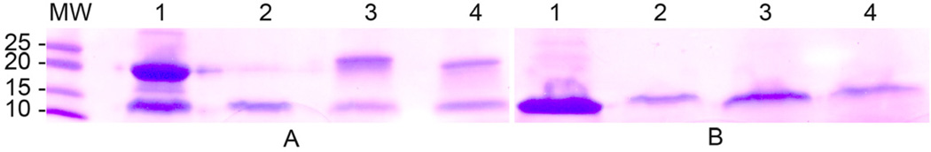
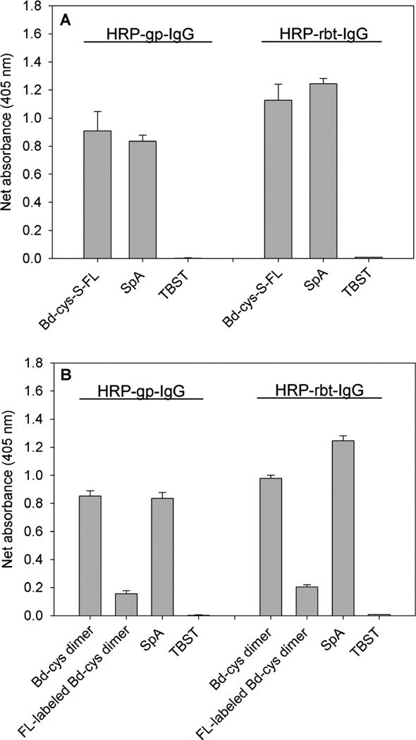
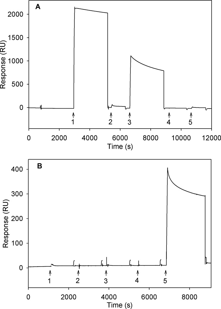
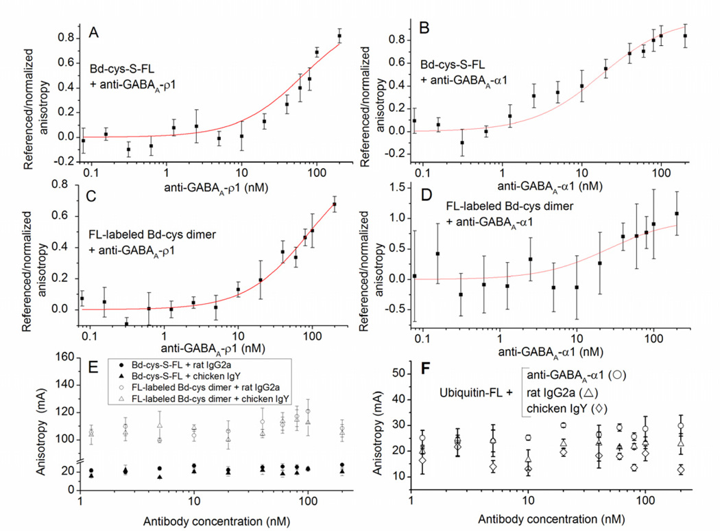
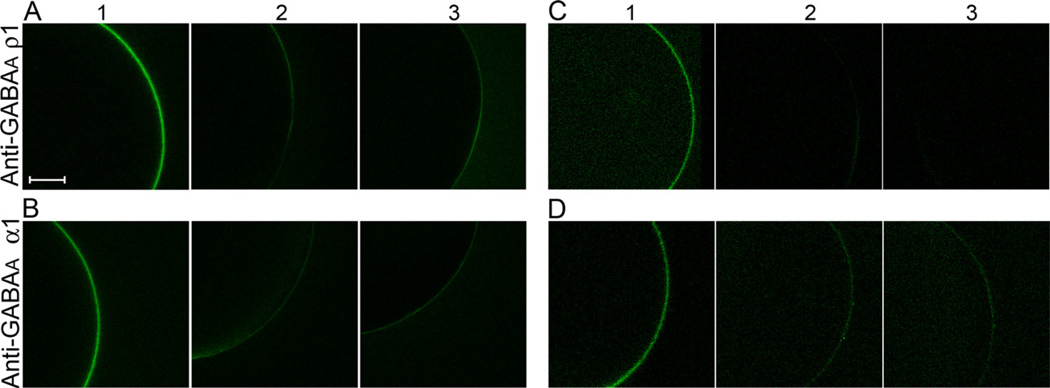
This study reports the preparation and characterization of cysteine-terminated B-domain (Bd-cys) of Staphylococcus aureus protein A, in combination with immunoglobulin G (IgG) antibodies directed against the ρ1 and α1 subunits of GABA(A) receptors, for localizing reagents of interest to the target receptor. A cysteine residue was inserted at the C terminus of the cysteine-lacking B-domain (Bd) and used for conjugating maleimide-containing compounds. As determined by enzyme-linked immunosorbent assay (ELISA), binding of a Bd-cys-S-fluorescein conjugate to polyclonal guinea pig anti-GABA(A)-ρ1 and rabbit anti-GABA(A)-α1 IgG was similar to that exhibited by full-length protein A. Surface plasmon resonance analysis of the interaction of Bd-cys-S-PEG3400-biotin conjugate (where PEG is polyethylene glycol) with anti-GABA(A)-ρ1 and anti-GABA(A)-α1 yielded K(D) values of 6.4 ± 1.9 and 0.4 ± 0.1 nM, respectively. Fluorescence anisotropy analysis of the binding of Bd-cys-S-fluorescein to the two antibodies yielded EC50 values of 65 and 18 nM, respectively. As determined with biotin-reactive fluorescent reagents, Bd-cys-S-PEG3400-biotin specifically bound to the plasma membrane of Xenopus laevis oocytes that expressed α1β2γ2 or homomeric ρ1 GABA(A) receptors and were pretreated with the corresponding anti-GABA(A) IgG. The IgG-binding specificity and high affinity of Bd-cys conjugates illustrate the potential of these conjugates, in combination with a selected IgG, to localize compounds of interest at specific cell surface proteins.


Figures

Fig. 1
Fig. 1. SDS-PAGE analysis
Profiles obtained for (1) affinity-purified Bd-cys, (2) Bd-cys-S-FL, (3) FL-labeled Bd-cys dimer, and (4) Bd-cys-S-PEG3400-biotin. Samples were prepared in buffer lacking β-ME (A) or containing 5% (v/v) β-ME (B) immediately prior to electrophoresis (4–20% acrylamide Tris-HCl gel). MW: molecular weight (kDa) of standards. Dimer bands were present in the profiles of all three conjugates (lanes 2–4) in non-reducing conditions (A), but not when the loading buffer contained β-ME (B).
Fig. 2
Fig. 2. ELISA for binding of Bd-cys preparations and SpA to HRP-conjugated guinea pig IgG (HRP-gp-IgG) and HRP-conjugated rabbit IgG (HRP-rbt-IgG)
Net absorbance (sample minus blank) data obtained at 405 nm, indicated as the mean ± SD of triplicate values. A: Bd-cys-S-FL and SpA binding vs. TBST control. B: Bd-cys dimer, FL-labeled Bd-cys dimer and SpA vs. TBST control. For both of the investigated IgGs, net absorbance obtained with the Bd-cys and SpA preparations in all cases exceeded that exhibited by the corresponding TBST control (p < 9.0 × 10⁻⁴).
Fig. 3
Fig. 3. SPR sensograms (responses in RU) for binding of IgG and IgY antibodies
A: Bd-cys-S-PEG3400-biotin capture agent. Here and in Panel B, arrows indicate the times of introduction of (1) anti-GABAA-α1 rabbit IgG, (2) rat IgG2a, (3) anti-GABAA-ρ1 guinea pig IgG, (4) chicken IgY, and (5) anti-insulin chicken IgY. B: Insulin-biotin capture agent. In both A and B, sensogram electronic spikes of ≤ 4 s duration have been removed. Surface-bound Bd-cys-S-PEG3400-biotin thus specifically binds anti-GABAA-α1 IgG and anti-GABAA-ρ1 IgG.
Fig. 4
Fig. 4. SPR sensograms (RU) for kinetics of binding of anti-GABAA-ρ1 and anti-GABAA-α1 to immobilized Bd-cys-S-PEG3400-biotin
A: Red: sensograms obtained with anti-GABAA-ρ1 at 20, 10, 5, 2.5, 1.25, and 0.625 nM. Black: fitted curves obtained using Biacore software in a 1:1 binding analysis. B: Sensograms (red) and fitted curves (black) obtained with the addition of anti-GABAA- α1 at 10, 5, 2.5, 1.25 and 0.625. nM. Binding kinetics of both antibodies to Bd-cys-S-PEG3400-biotin are well described by Langmuir 1:1 binding analysis (see Methods).
Fig. 5
Fig. 5. Binding of Bd-cys preparations to anti-GABAA-subunit antibodies determined by FA
The horizontal axis in each panel represents concentration of the tested antibody. (A–D) Referenced/normalized anisotropy in relation to antibody concentration (mean ± SD of values obtained in ≥ 2 experiments, each involving duplicate/triplicate samples). A: Bd-cys-S-FL with anti-GABAA-ρ1. Here and in (B–D), curves illustrate equation (1) fitted to the data. B: Bd-cys-S-FL with anti-GABAA-α1. C: FL-labeled Bd-cys dimer with anti-GABAA-ρ1. D: FL-labeled Bd-cys dimer with anti-GABAA-α1. (E–F) Anisotropy (mA) in relation to antibody concentration (mean ± SD of values obtained in one experiment, each involving triplicate samples). E: Anisotropy (mA) for Bd-cys preparations with control antibodies. Mixtures were: Bd-cys-S-FL with rat IgG2a (filled circles); Bd-cys-S-FL with chicken IgY (filled triangles); FL-labeled Bd-cys dimer with rat IgG2a (open circles); and FL-labeled Bd-cys dimer with chicken IgY (open triangles). F: Anisotropy (mA) for ubiquitin-FL with anti-GABAA-α1 (open circles), rat IgG2a (open triangles) and chicken IgY (open diamonds). An increase in anisotropy with antibody concentration occurred only when the antibody incubated with the Bd preparation was anti-GABAA-ρ1 or anti-GABAA-α1.
Fig. 6
Fig. 6. Binding of Bd-cys preparations and anti-GABAA-subunit antibodies to GABAA-expressing oocytes
A: GABAA-ρ1-expressing oocyte with anti-GABAA-ρ1, Bd-cys-S-PEG3400-biotin and streptavidin-DyLight 488 (column 1) or with Bd-cys-S-PEG3400-biotin and streptavidin-DyLight 488 only (column 2). Column 3: non-expressing oocyte with same treatment as in column 1. B: α1β2γ2 GABAA-expressing oocyte with anti-GABAA-α1, Bd-cys-S-PEG3400-biotin and streptavidin-DyLight 488 (column 1) or with Bd-cys-S-PEG3400-biotin and streptavidin-DyLight 488 only (column 2). Column 3: non-expressing oocyte with same treatment as in column 1. C: GABAA-ρ1-expressing oocyte with anti-GABAA-ρ1 and FL-labeled Bd-cys dimer (column 1) or with FL-labeled Bd-cys dimer only (column 2). Column 3: non-expressing oocyte with same treatment as in column 1. D: α1β2γ2 GABAA-expressing oocyte with anti-GABAA-α1 and FL-labeled Bd-cys dimer (column 1) or with FL-labeled Bd-cys dimer only (column 2). Column 3: non-expressing oocyte with same treatment as in column 1. Similar microscope acquisition settings were used for all conditions. For visual clarity, a fixed adjustment of brightness and contrast was applied to all images of C and D. Scale bar = 150 µm. The data show that the binding of Bd-cys-S-PEG3400-biotin (A-B) and of FL-labeled Bd-cys dimer (C-D) to the oocyte surface membrane depends on both the expression of GABAA receptor (GABAA-ρ1 in A and C; α1β2γ2 GABAA in B and D) and the presence of the cognate antibody.

Similar articles

Cited by

References

    1. Graille M, Stura EA, Corper AL, Sutton BJ, Taussig MJ, Charbonnier J-B, Silverman GJ. Crystal structure of a Staphylococcus aureus protein A domain complexed with the Fab fragment of a human IgM antibody: structural basis for recognition of B-cell receptors and superantigen activity. Proc. Natl. Acad. Sci. U.S.A. 2000;97:5399–5404. - PMC - PubMed
    1. Cedergren L, Andersson R, Jansson B, Uhlén M, Nilsson B. Mutational analysis of the interaction between staphylococcal protein A and human IgG1. Protein Eng. 1993;6:441–448. - PubMed
    1. Langone JJ. 125I-Labeled protein A: reactivity with IgG and use as a tracer in radioimmunoassay. Methods Enzymol. 1980;70(A):356–375. - PubMed
    1. MacSween JM, Eastwood SL. Recovery of antigen from Staphylococcal protein A-antibody adsorbents. Methods Enzymol. 1981;73:459–471. - PubMed
    1. Ljungberg UK, Jansson B, Niss U, Nilsson R, Sandberg BE, Nilsson B. The interaction between different domains of staphylococcal protein A and human polyclonal IgG, IgA, IgM, and F(ab’)2: separation of affinity from specificity. Mol. Immunol. 1993;30:1279–1285. - PubMed

Publication types

LinkOut - more resources