. 2012 Nov 7;20(11):1905-17.
doi: 10.1016/j.str.2012.08.025. Epub 2012 Sep 20.

The structure of the plk4 cryptic polo box reveals two tandem polo boxes required for centriole duplication

Affiliations

The structure of the plk4 cryptic polo box reveals two tandem polo boxes required for centriole duplication

Lauren K Slevin et al. Structure. .

Abstract

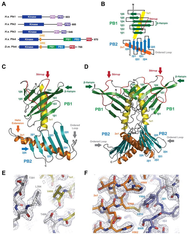
Centrioles are key microtubule polarity determinants. Centriole duplication is tightly controlled to prevent cells from developing multipolar spindles, a situation that promotes chromosomal instability. A conserved component in the duplication pathway is Plk4, a polo kinase family member that localizes to centrioles in M/G1. To limit centriole duplication, Plk4 levels are controlled through trans-autophosphorylation that primes ubiquitination. In contrast to Plks 1-3, Plk4 possesses a unique central region called the "cryptic polo box." Here, we present the crystal structure of this region at 2.3 Å resolution. Surprisingly, the structure reveals two tandem homodimerized polo boxes, PB1-PB2, that form a unique winged architecture. The full PB1-PB2 cassette is required for binding the centriolar protein Asterless as well as robust centriole targeting. Thus, with its C-terminal polo box (PB3), Plk4 has a triple polo box architecture that facilitates oligomerization, targeting, and promotes trans-autophosphorylation, limiting centriole duplication to once per cell cycle.


Figures

Figure 1
Figure 1
The Plk4 CPB is Composed of Tandem PB domains, PB1 and PB2. A) Polo-like kinase family domain architecture. Plk1-3 contain two PB domains, Plk4 contains three PB domains. Plk4 levels are regulated by the DRE (orange). B) Secondary structure topology diagram of Plk4’s conserved central domain: PB1 (βstrands in green, α-helix in yellow, stirrup in red, loops in black) and PB2 (β-strands in blue, α-helix in orange, loops in black). C) Tertiary structure of the Plk4 PB1-PB2 monomer colored as in B. D) Quaternary structure of homodimeric Plk4 PB1-PB2, rotated 90° relative to C. E)Stick representation of the junction between PB1 1α1 and PB2 where the T449 hydroxyl caps the 1α1 helix. F) Stick representation of the PB2 intra-domain disulfide formed between 2α1 C566 and 2β2 C511. Final 2Fo-Fc electron density contoured at 1.5σ (E,F).
Figure 2
Figure 2
Plk4 PB1-PB2 is an Asymmetric Homodimer with Plastic Stirrups and Loops. A) Superposition of Plk4 PB1-PB2 protomers A and B showing plasticity in the stirrup as well as the 2β4-2β5 and 2β5-2α1 loops. Plk4 colored as in Figure 1B, shown in cartoon format. B) Plk4 PB1-PB2 protomer A backbone colored and scaled according to Cα B-factor values ranging from 14 (dark blue) to 105 (red) Å2. High B-factors correlate with the structurally plastic loop segments between protomers A and B (shown in A). The region bridging PB1 and PB2 shows little structural divergence and is dominated by low B-factor values. Plk4 PB1-PB2 protomer A (C), and protomer B (D) shown in sphere format; oriented as shown in the inset. Residues involved in homodimerization from both protomers, unique to protomer A, and unique to protomer B, are colored dark grey, purple, and raspberry, respectively. E) Size exclusion chromatography – multi-angle light scattering analysis of H6-Plk4 PB1-PB2 injected at 19 μM (red trace) and 28 μM (green trace)(100 μl). Y-axis at left displays molecular weight (kDa), Y-axis at right displays normalized differential refractive index, X-axis displays time component of the run. (See Figure S1).
Figure 3
Figure 3
Plk4 PB1 and PB2 Diverge from Plk1 PB Domain Structures and Form a Unique Inter-domain Interaction. A) Structural alignment of Plk4 PB1 and Plk4 PB2 (colored as in Figure 1B) with human Plk1 PB1 bound to a phosphopeptide target (3FVH, β-strands in purple, α-helix in grey, peptide in blue). B) Matrix showing the rmsd (Å) between structures of human Plk1 PB1 and PB2, fly Plk4 PB1 and PB2, and mouse Plk4 PB3. C) Superposition of Plk4 PB1-PB2 homodimer and the Plk1 PB1-PB2-phosphopeptide structure, aligned over Plk4 PB1 and Plk1 PB1, highlighting the differential organization of Plk1 PB1-PB2 as compared to Plk4 PB1-PB2. The Plk4 PB1 stirrup overlaps with the corresponding binding site of Plk1 PB2. The location where Plk1 PB1 binds its phosphopeptide target is accessible on Plk4 PB1. D) Superposition of Plk4 PB1-PB2 and the Plk4 PB3 homodimer, aligned over single PB1 and PB3 domains. Insets show the orientation of each independent structure (C,D).
Figure 4
Figure 4
Plk4 PB1 and PB2 Form a Composite Inter-domain Groove Delineated by Conserved and Basic Residues. A) Plk4 sequence alignment across ten species. Protomer A solvent accessible surface area (ASA) (Å2) is indicated. 100% identity is highlighted in green, 80% identity in yellow (homologous residues also highlighted in yellow where the 80% identity criteria is met). Human Plk1 PB1 sequence is aligned against Plk4 PB1 and PB2, based on structural alignment. Residues involved in homodimerization are indicated below the alignment, colored as in Fig 2C,D. B) Plk4 PB1-PB2 homodimer structure shown in sphere format with conserved residues colored as in A (left) and in surface representation (right) showing electrostatics contoured from −2.0 to +2.0 kT/e. C) Protomer A rotated 45° relative to the orientation shown in B showing conservation and electrostatics as in B. The phosphopeptide from the Plk1 PB1-PB2 structure (3FVH) shown in stick format and colored blue, is docked onto the Plk4 structure in both B and C, based on the structural alignment of Plk1 PB1 and Plk4 PB1 shown in Fig 3C. D–E) Anti-GFP immunoprecipitates from S2 cell lysates transiently-expressing N-terminal Asl-V5 and the indicated Plk4-GFP construct or control GFP, probed for GFP and V5.
Figure 5
Figure 5
The Plk4 PB1-PB2 Cassette is Required for Robust Centriole Localization.A) Schematic of PB-GFP containing constructs assayed for centriole localization. B) S2 cells were transiently-transfected with the constructs shown in A (green), induced to express for 24 hours, and immunostained with anti D-PLP antibody to mark centrioles (red). Centriole co-localization was classified as strong, weak, or no co-localization. Total number of cells analyzed and independent experiments performed noted at right. C, D) Expression of PB1-PB3 (B) or the PB1-PB2 cassette (D) results in strong centriole co-localization. Cell margins are indicated (white dashed lines). E–I) Single or incomplete PBs primarily display weak or no centriole co-localization. Representative images of differential localization are shown. Boxed regions are magnified in the lower panels. Scale, 5μm.
Figure 6
Figure 6
Plk4 PB1-PB2 Promotes Centriole Amplification and Protects Full-length Plk4 in trans. A) S2 cells were transiently-transfected with either inducible GFP, Plk4 PB1-PB2-GFP, or non-degradable Plk4-SBM-GFP (green), induced for 3 days, fixed, and stained for centrioles (PLP, red) and DNA (blue). Arrowheads mark centrioles. Boxed regions are magnified in the insets and highlight centriole clusters not observed in controls. B) Histograms of centriole counts were measured from S2 cells transiently expressing the indicated constructs after 3 days of induction (see Figures S3,4). The percentage of cells with a centriole count per cell <2, 2, and >2 is indicated. Error bars indicate standard deviation. C) Ectopic Plk4 PB1-PB2-GFP expression is sufficient to stabilize full-length Plk4-GFP. Immunoblots of S2 cell lysates showing that overexpression of PB1-PB2-GFP (but not Plk4-Δ[PB1-PB2]-GFP or GFP) stabilizes wild-type full-length Plk4-GFP. Tubulin, loading control. D) PB1-PB2-GFP co-immunoprecipitates Plk4-FL-myc. Immunoblots of anti-GFP immunoprecipitates from S2 cells co-transfeted with Plk4-FL-myc and PB1-PB2-GFP or GFP (control). E) Anti-GFP immunoblot of Plk4-GFP constructs transiently expressed in S2 cells showing differential stability. Tubulin, loading control. F) Plk4-FL-GFP is expressed but rapidly degraded by Slimb-mediated ubiquitination. Cell lysates from S2 cells transfected with Plk4-FL-GFP and treated with control or Slimb dsRNA. Tubulin, loading control. G) Plk4 lacking the PB1-PB2 cassette shows reduced auto-phosphorylation of the DRE as assayed by Slimb binding. Immunoblots of anti-GFP immunoprecipitates from S2 cells transfected with Plk4-FL-GFP or Plk4-Δ[PB1-PB2]-GFP and blotted for GFP and Slimb. H) Model of the Plk4 homodimer. PB1 and PB2 mediate homodimerization. PB1 and PB2 form a composite Asl/Cep152 binding site, recruiting Plk4 to the centriole. PB1-PB2 homodimerization scaffolds Plk4 trans-autophosphorylation, priming the DRE for SCFSlimb binding and ubiquitination.

Comment in

References

    1. Adams PD, Afonine PV, Bunkóczi G, Chen VB, Davis IW, Echols N, Headd JJ, Hung LW, Kapral GJ, Grosse-Kunstleve, et al. PHENIX: a comprehensive Python-based system for macromolecular structure solution. Acta Cryst. 2010;D66(Part A):213–221. - PMC - PubMed
    1. Archambault V, Glover DM. Polo-like kinases: conservation and divergence in their functions and regulation. Nat Rev Mol Cell Biol. 2009;10:265–275. - PubMed
    1. Azimzadeh J, Marshall WF. Building the centriole. Curr Biol. 2010;20:816–825. - PMC - PubMed
    1. Bettencourt-Dias M, Hildebrandt F, Pellman D, Woods G, Godinho SA. Centrosomes and cilia in human disease. Trends Genet. 2011;27:307–315. - PMC - PubMed
    1. Bettencourt-Dias M, Rodrigues-Martins A, Carpenter L, Riparbelli M, Lehmann L, Gatt MK, Carmo N, Balloux F, Callaini G, Glover DM. SAK/PLK4 is required for centriole duplication and flagella development. Curr Biol. 2005;15:2199–2207. - PubMed

Publication types

Substances