Comparative Study
. 2012 Sep;13(3):299-310.
doi: 10.4142/jvs.2012.13.3.299.

Comparing the osteogenic potential of canine mesenchymal stem cells derived from adipose tissues, bone marrow, umbilical cord blood, and Wharton's jelly for treating bone defects

Affiliations
Comparative Study

Comparing the osteogenic potential of canine mesenchymal stem cells derived from adipose tissues, bone marrow, umbilical cord blood, and Wharton's jelly for treating bone defects

Byung-Jae Kang et al. J Vet Sci. 2012 Sep.

Abstract

Alternative sources of mesenchymal stem cells (MSCs) for replacing bone marrow (BM) have been extensively investigated in the field of bone tissue engineering. The purpose of this study was to compare the osteogenic potential of canine MSCs derived from adipose tissue (AT), BM, umbilical cord blood (UCB), and Wharton's jelly (WJ) using in vitro culture techniques and in vivo orthotopic implantation assays. After canine MSCs were isolated from various tissues, the proliferation and osteogenic potential along with vascular endothelial growth factor (VEGF) production were measured and compared in vitro. For the in vivo assay, MSCs derived from each type of tissue were mixed with β-tricalcium phosphate and implanted into segmental bone defects in dogs. Among the different types of MSCs, AT-MSCs had a higher proliferation potential and BM-MSCs produced the most VEGF. AT-MSCs and UCB-MSCs showed greater in vitro osteogenic potential compared to the other cells. Radiographic and histological analyses showed that all tested MSCs had similar osteogenic capacities, and the level of new bone formation was much higher with implants containing MSCs than cell-free implants. These results indicate that AT-MSCs, UCB-MSCs, and WJ-MSCs can potentially be used in place of BM-MSCs for clinical bone engineering procedures.


Figures

Fig. 1
Fig. 1
The orthotopic implantation procedure. (A) β-tricalcium phosphate (β-TCP) mixed with canine mesenchymal stem cells (MSCs). (B) Segmental defect in the radial diaphysis. (C) Filling the bone defect with β-TCP compound and MSCs. (D) Complete implantation. (E) Implant harvested after 20 weeks.
Fig. 2
Fig. 2
Morphologic comparison and fluorescence-activated cell sorting (FACS) analysis of various cultured canine MSCs at passage 3. MSCs were isolated from different tissues including (A) adipose tissue (AT), (B) bone marrow (BM), (C) umbilical cord blood (UCB) and (D) Wharton's jelly (WJ). All the cells had a typical fibroblast-like morphology. (E and F) FACS analysis revealed that AT-MSCs, BM-MSCs, UCB-MSCs, and WJ-MSCs expressed CD44, CD73, CD90, and CD105 but not CD14, CD34, or CD45. A~D: ×40. FITC: fluorescein isothiocyanate, PE: phycoerythrin.
Fig. 3
Fig. 3
Cumulative population doubling levels of canine MSCs derived from various tissues. Population doubling was measured at each passage. Data are expressed as the mean ± SD (n = 3).
Fig. 4
Fig. 4
Mineralization assay results and alkaline phosphatase (ALP) activity of the various MSCs. (A) AT-MSCs (a1 and a2), BM-MSCs (b1 and b2), UCB-MSCs (c1 and c2), and WJ-MSCs (d1 and d2) were seeded and cultured in osteogenic medium for 2 weeks after the cells reached confluence. To confirm the presence of calcium deposits, cells were stained with Alizarin Red S. Quantification of mineralization (B) and ALP activity (C) was performed to compare in vitro osteogenic capabilities of the MSCs. Data are presented as the mean ± SD (n = 3). *Indicates a statistically significant difference (p < 0.05) compared to the AT-MSCs under the same conditions. Indicates a statistically significant difference (p < 0.05) compared to the UCB-MSCs. a2, b2, c2, and d2: ×40.
Fig. 5
Fig. 5
Quantification of vascular endothelial growth factor (VEGF) produced by the various MSCs. Data are presented as the mean ± SD (n = 3). *Indicates a statistically significant difference (p < 0.05) compared to the control under the same conditions. Indicates a statistically significant difference (p < 0.05) compared to the AT-MSCs. Indicates a statistically significant difference (p < 0.05) compared to the BM-MSCs.
Fig. 6
Fig. 6
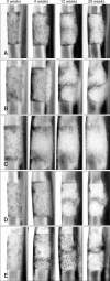
Mediolateral radiographs of the treated defects obtained immediately after the operation as well as 4, 12, and 20 weeks after surgery. The radiolucent transverse zone at the defect ends remained distinct in the control group (A) after 20 weeks. In contrast, union at the host bone-implant interfaces was evident in the AT-MSC (B), BM-MSC (C), UCB-MSC (D), and WJ-MSC (E) groups.
Fig. 7
Fig. 7
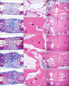
Coronal sections of the segmental bone defects 20 weeks after implantation. Macroscopic views of the repaired area in each group are shown: (A) control, (B) AT-MSC, (C) BM-MSC, (D) UCB-MSC, and (E) WJ-MSC groups. The proximal portion is at the left of the panels (A~E). Bone was stained purple and residual β-TCP was light blue. Bone formation was observed on the surface the β-TCP throughout the defect in all groups. However, there was a higher amount of bone formation in all experimental groups compared to the control animals. Detailed views of the inside (A1, B1, C1, D1, and E1) and interfacial (A2, B2, C2, D2, and E2) areas are shown. In the control group, a minimal amount of new bone was observed in the inside area (A1), and cortical continuity was not found at the interfacial area (A2). In the experimental groups, osteocytes (arrow) and hematopoietic tissues (arrow head) embedded in substantial bone matrix were found together with some intact β-TCP particles in the inside area (B1, C1, D1, and E1). Cortical bony bridging had also formed at the interfacial area (B2, C2, D2, and E2). AZAN (A~E) and H&E (A1, 2, B1, 2, C1, 2, D1, 2, E1, and 2) stain. A1, B1, C1, D1, and E1: ×200, A2, B2, C2, D2, and E2: ×40. b: bone, s: β-TCP scaffold, ncb: native cortical bone area, nba: newly formed bone area.

References

    1. Al-Khaldi A, Eliopoulos N, Martineau D, Lejeune L, Lachapelle K, Galipeau J. Postnatal bone marrow stromal cells elicit a potent VEGF-dependent neoangiogenic response in vivo. Gene Ther. 2003;10:621–629. - PubMed
    1. Arinzeh TL, Peter SJ, Archambault MP, van den Bos C, Gordon S, Kraus K, Smith A, Kadiyala S. Allogeneic mesenchymal stem cells regenerate bone in a critical-sized canine segmental defect. J Bone Joint Surg Am. 2003;85:1927–1935. - PubMed
    1. Arthur A, Zannettino A, Gronthos S. The therapeutic applications of multipotential mesenchymal/stromal stem cells in skeletal tissue repair. J Cell Physiol. 2009;218:237–245. - PubMed
    1. Bartholomew A, Sturgeon C, Siatskas M, Ferrer K, McIntosh K, Patil S, Hardy W, Devine S, Ucker D, Deans R, Moseley A, Hoffman R. Mesenchymal stem cells suppress lymphocyte proliferation in vitro and prolong skin graft survival in vivo. Exp Hematol. 2002;30:42–48. - PubMed
    1. Bianco P, Kuznetsov SA, Riminucci M, Robey PG. Postnatal skeletal stem cells. Methods Enzymol. 2006;419:117–148. - PubMed

Publication types

MeSH terms