Review
. 2012 Dec;143(6):1442-1460.e1.
doi: 10.1053/j.gastro.2012.09.032. Epub 2012 Sep 20.

Epigenetics of colorectal cancer

Affiliations
Review

Epigenetics of colorectal cancer

Ajay Goel et al. Gastroenterology. 2012 Dec.

Abstract

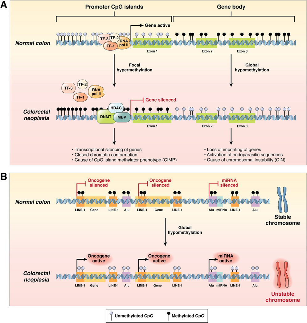
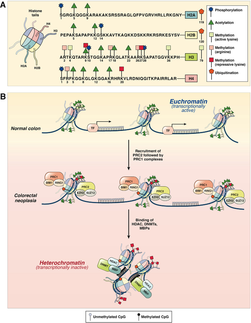
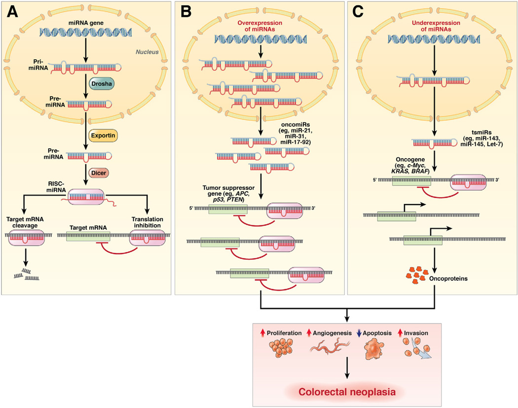
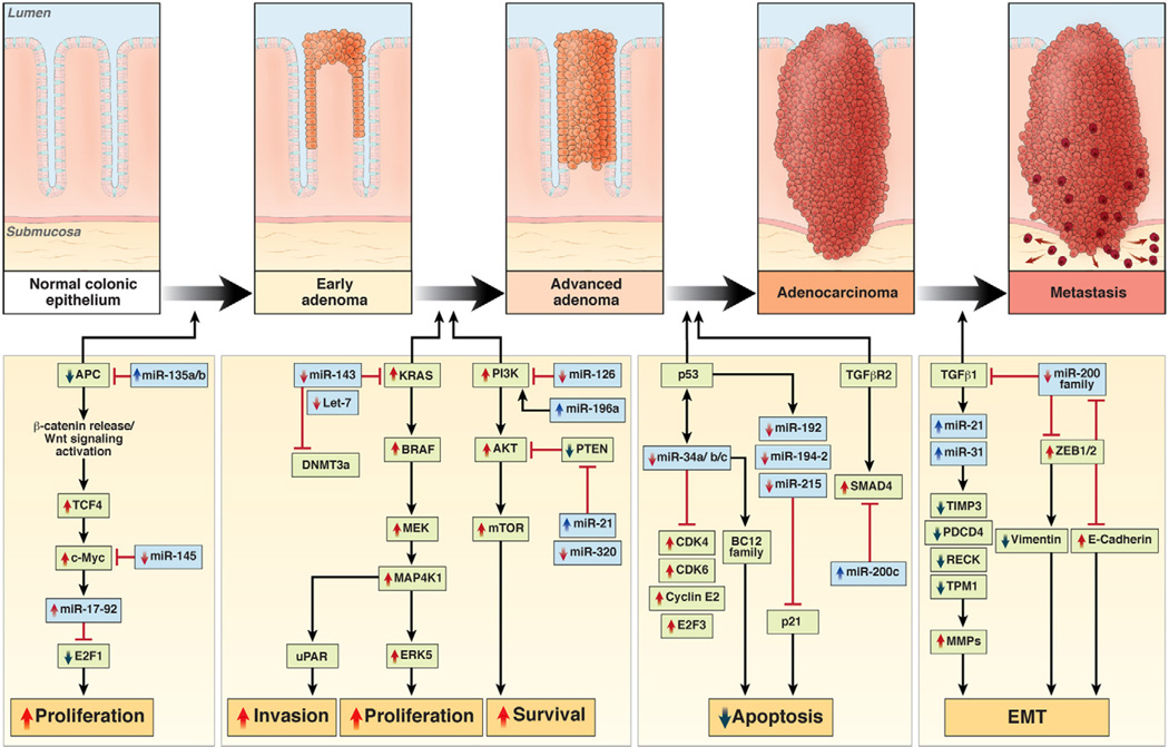
In the early years of the molecular biology revolution, cancer research was mainly focused on genetic changes (ie, those that altered DNA sequences). Although this has been extremely useful as our understanding of the pathogenesis and biology of cancer has grown and matured, there is another realm in tumor development that does not involve changing the sequence of cellular DNA. This field is called "epigenetics" and broadly encompasses changes in the methylation of cytosines in DNA, changes in histone and chromatin structure, and alterations in the expression of microRNAs, which control the stability of many messenger RNAs and serve as "master regulators" of gene expression. This review focuses on the epigenetics of colorectal cancer and illustrates the impact epigenetics has had on this field.


Conflict of interest statement

Conflicts of interest

The authors disclose no conflicts.

Figures

Figure 1
Figure 1
DNA methylation patterns in the normal colon and colorectal neoplasia. (A) The process of DNA hypermethylation-induced transcriptional silencing of tumor suppressor genes in colorectal neoplasia vis-à-vis normal colonic cells. In this instance, altered DNA methylation occurs at CpG dinucleotides either in the context of CpG-rich “promoter CpG islands” or at the abundant but sparsely distributed CpG sites throughout the “gene body.” In normal cells, CpG sites within the 5′ promoter CpG islands upstream of the transcription start site of a tumor suppressor gene are generally unmethylated (blue circles), whereas the dinucleotide repeats within the gene body are frequently methylated (black circles). This configuration permits easy and uninterrupted access by transcription factors (eg, TF-1, TF-2, TF-3) and RNA polymerase II (RNA pol II) to bind to the gene promoter and facilitate active gene expression. In contrast, in colorectal neoplasia, DNMT in association with HDAC and methyl binding proteins (MBP) catalyze the transfer of methyl groups to cytosines in the promoter CpG sites, resulting in the hypermethylation-induced transcriptional silencing of the associated tumor suppressor gene, which reflects CIMP. Concurrent with hypermethylation of gene promoters, CpG sites within the gene body experience DNA hypomethylation, which causes loss of imprinting of genes, activation of endoparasitic sequences, and chromosomal instability in CRC cells. (B) DNA hypomethylation may also occur at repetitive DNA sequences and within the promoter regions of miRNAs in colorectal neoplasia compared with normal colonic cells (see text). Evolutionarily conserved proto-oncogenes (indicated as “Gene” in the figure) and oncogenic miRNAs (indicated as “miRNA” in the figure) are not expressed in healthy normal colonic cells. Expression of these oncogenes and miRNAs is inhibited by hypermethylation of the promoter CpG sites that occurs within LINE-1 and Alu repeat elements. However, in colorectal neoplasia, LINE-1 and Alu sequences become hypomethylated, which permits the potential activation of previously silenced proto-oncogenes and oncogenic miRNAs as well as chromosomal instability.
Figure 2
Figure 2
(A) Histone modification patterns in colorectal neoplasia. The 4 histone proteins (H2A, H2B, H3, and H4), each with 2 copies, associate in cylindrical structures that constitute the histone core, and each nucleosome is a chromatin unit composed of 150 to 200 base pairs of DNA tightly wrapped around the octameric histone core. Covalent modifications of each histone tail, including acetylation, methylation, phosphorylation, and ubiquitination, are also shown on the right. The amino acid sequences of the N-terminal tails and the key selected sites of modification of these 4 histones are shown. (B) In normal colon, histones are arranged in the open, euchromatin configuration, which represents transcriptionally active chromatin (upper panel). Histone tails may undergo multiple modifications, which include histone acetylation (green triangles) and lysine methylation (green squares). Gene promoters in euchromatin allow easy access for transcription factor (TF) binding, permitting active gene expression. In CRC cells, chromatin configuration is converted to the more compacted heterochromatin configuration, which is transcriptionally inactive and results in the hypermethylation-induced silencing of genes. This process is facilitated by the multimeric polycomb repressive protein complexes, PRC2 and PRC1, which sequentially bind to histones and initiate histone methylation (initiated by PRC2 complex containing EZH2 and SUZ12) followed by maintenance methylation and monoubiquitination (performed by PRC1 complex with BIM1 and RING1 proteins). This is followed by the recruitment of DNMTs, HDACs, and methyl binding proteins (MBPs) to complete the PRC-mediated transcriptional silencing of genes.
Figure 3
Figure 3
miRNA genesis in normal colon and their role as oncogenic and tumor suppressive genes in colorectal neoplasia. (A) In normal cells, primary miRNA transcripts are processed into precursor miRNAs by an enzymatic complex that includes the nuclear ribonuclease III enzyme Drosha. The resulting precursor miRNA is transported to the cytoplasm by exportin, where it is processed into ~22-nucleotide duplexes by the ribonuclease III enzyme Dicer. The strand corresponding to the mature miRNA is subsequently loaded onto the RNA-induced silencing complex (RISC). Mature miRNAs bind to the 3′ untranslated regions of target mRNAs and can suppress their expression through translational inhibition, degradation of the target miRNA, or both. (B) In cancer cells, miRNAs can act as oncogenes (or oncomiRs) and can be overexpressed during different stages of cancer development. Overexpression of specific oncomiRs in colorectal adenomas and cancers (eg, miR-21, −31, −17, −92) results in targeting and suppression of their target tumor suppressor genes (eg, APC, p53, PTEN). (C) Likewise, miRNAs may act as tumor suppressors (or tsmiRs), in which these miRNAs are down-regulated in CRC cells (eg, miR-143, -145, let-7) and are unable to block the expression of their target oncogenes completely (eg, c-Myc, KRAS, BRAF). The overall consequence of oncomiR and tsmiR regulation in cancer cells might involve increased proliferation, increased invasiveness or angiogenesis, and decreased apoptosis, all of which facilitate the development of colorectal neoplasia.
Figure 4
Figure 4
Involvement of miRNAs in multistep colorectal carcinogenesis. Various miRNAs have been identified that are dysregulated in colorectal neoplasia (in blue), along with validated gene targets (in green) in the multistep Vogelgram. Although the number of newly discovered miRNAs and gene targets is continuously growing, this figure provides evidence that miRNAs can regulate any of the major pathways in colorectal neoplasia, including their ability to regulate β-catenin/WNT signaling, MAPK and p53 signaling, proliferation, apoptosis, cell cycle control, migration, and invasion.

References

    1. Gardiner-Garden M, Frommer M. CpG islands in vertebrate genomes. J Mol Biol. 1987;196:261–282. - PubMed
    1. Lao VV, Grady WM. Epigenetics and colorectal cancer. Nat Rev Gastroenterol Hepatol. 2011;8:686–700. - PMC - PubMed
    1. Graff JR, Herman JG, Myohanen S, et al. Mapping patterns of CpG island methylation in normal and neoplastic cells implicates both upstream and downstream regions in de novo methylation. J Biol Chem. 1997;272:22322–22329. - PubMed
    1. Ooi SL, Henikoff S. Germline histone dynamics and epigenetics. Curr Opin Cell Biol. 2007;19:257–265. - PubMed
    1. Chadwick BP, Willard HF. Chromatin of the Barr body: histone and non-histone proteins associated with or excluded from the inactive X chromosome. Hum Mol Genet. 2003;12:2167–2178. - PubMed

Publication types