. 2012 Sep;24(9):3684-94.
doi: 10.1105/tpc.112.099507. Epub 2012 Sep 21.

Two interacting proteins are necessary for the editing of the NdhD-1 site in Arabidopsis plastids

Affiliations

Two interacting proteins are necessary for the editing of the NdhD-1 site in Arabidopsis plastids

Clément Boussardon et al. Plant Cell. 2012 Sep.

Abstract

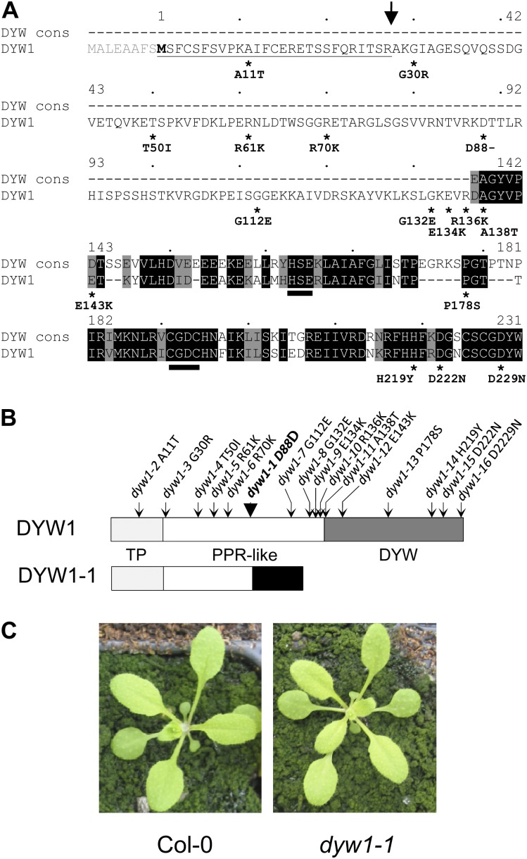
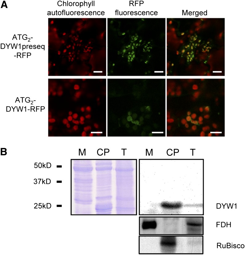
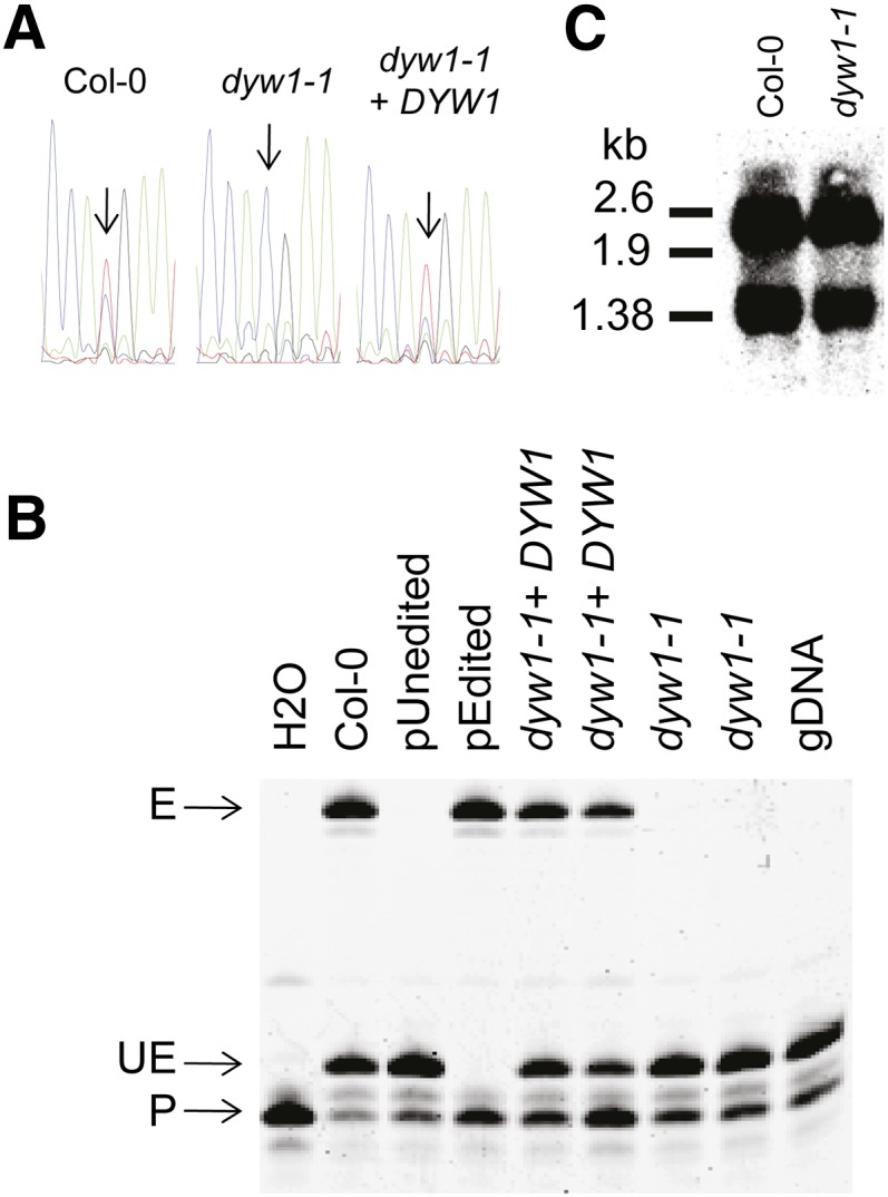
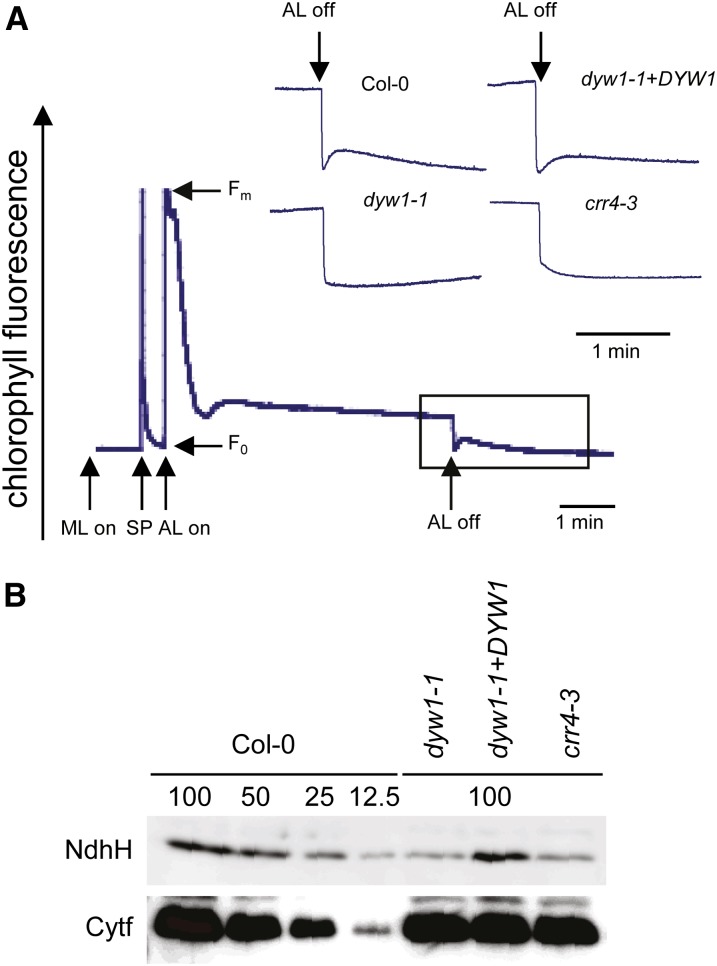
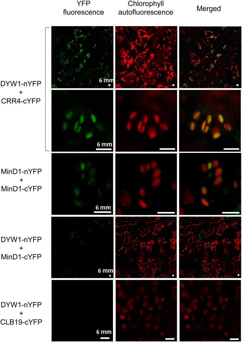
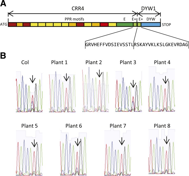
After transcription, mRNA editing in angiosperm chloroplasts and mitochondria results in the conversion of cytidine to uridine by deamination. Analysis of Arabidopsis thaliana mutants affected in RNA editing have shown that many pentatricopeptide repeat proteins (PPRs) are required for specific cytidine deamination events. PPR proteins have been shown to be sequence-specific RNA binding proteins allowing the recognition of the C to be edited. The C-terminal DYW domain present in many editing factors has been proposed to catalyze C deamination, as it shows sequence similarities with cytidine deaminases in other organisms. However, many editing factors, such as the first to be discovered, CHLORORESPIRATORY REDUCTION4 (CRR4), lack this domain, so its importance has been unclear. Using a reverse genetic approach, we identified DYW1, an RNA editing factor acting specifically on the plastid ndhD-1 editing site recognized by CRR4. Unlike other known editing factors, DYW1 contains no identifiable PPR motifs but does contain a clear DYW domain. We were able to show interaction between CRR4 and DYW1 by bimolecular fluorescence complementation and to reconstitute a functional chimeric CRR4-DYW1 protein complementing the crr4 dyw1double mutant. We propose that CRR4 and DYW1 act together to edit the ndhD-1 site.


Figures

Figure 1.
Figure 1.
Structure of the Wild-Type DYW1 Protein and the Mutant Proteins Encoded by the Different Alleles. (A) Alignment of the DYW1 sequence with the DYW domain consensus (Lurin et al., 2004). The DYW1 sequence was aligned against the DYW domain consensus (DYW cons) using the pairwise alignment software at http://pir.georgetown.edu/pirwww/search/pairwise.shtml. The sequence numbering is shown above the sequence according to M2. Identical residues are shaded black, and similar residues are shaded gray. The putative DYW1 targeting peptide is underlined, and the position of a potential cleavage site indicated by an arrow. The HxExnCxxC deaminase signature identified in DYW domains (Salone et al., 2007) is indicated by two black bars. Positions of the 16 substitutions identified in DYW1 coding sequence are indicated by stars under the DYW1 protein sequence. The sequence modification resulting from each mutation is indicated at the protein sequence level (amino acid identity in wild-type sequence/position of the amino acid in the protein sequence/ amino acid identity in the mutant protein). (B) Structure of the 231–amino acid protein encoded by the DYW1 gene and positions of the EMS mutations. The 27–amino acid targeting peptide (TP) is indicated in light gray and the 95–amino acid DYW domain in dark gray. The PPR-like region in the middle is indicated in white. The PPR-like region of DYW1 is not recognized by any PPR detection software but presents a low similarity to PPR proteins when searching by BLAST. Positions of the mutations are indicated by arrows on the DYW1 protein. The sequence modification resulting from each mutation is indicated at the protein sequence level (amino acid identity in wild-type sequence/position of the amino acid in the protein sequence/amino acid identity in the mutant protein). The structure of the putative DYW1-1 is indicated below the wild-type DYW1 protein. The additional protein sequence unrelated to wild-type DYW1 protein is indicated in black. (C) Phenotype of a dyw1-1 mutant and Col-0 wild-type plant after 18 d in soil. [See online article for color version of this figure.]
Figure 2.
Figure 2.
DYW1 Is Localized in Chloroplasts of Arabidopsis Cells. (A) Fluorescence images of Arabidopsis plantlets transiently expressing DYW1-RFP fusion proteins. Either the first 100–amino acid polypeptide or the full-length DYW1 protein, starting both at the M2 Met encoded by the ATG2 codon, were expressed as a fusion with the RFP at the C terminus. Plantlets were observed by confocal microscopy 4 d after transformation. Bars = 10 μm. (B) Immunoblot analysis of total extract (T), mitochondrial (M), and chloroplast (CP) protein fractions with antibodies directed against DYW1. Protein fractions from Arabidopsis were analyzed by immunoblots. Loading control (Coomassie blue staining; left panel) shows that equal amounts of total protein were loaded. Black bars represent molecular mass marker positions. The purity of the two fractions was tested with antibodies directed against the mitochondrial formate dehydrogenase (FDH) and the chloroplast large subunit of ribulose-1,5-bisphosphate carboxylase/oxygenase (RuBisCo).
Figure 3.
Figure 3.
The dyw1-1 Mutant Lacks Editing of the ndhD-1 Site. (A) Nucleotide sequences of RT-PCR products obtained from Col-0, dyw1-1, and complemented dyw1-1 cDNA are shown as sequencing chromatograms. An arrow pointing to the corresponding peak indicates the ndhD-1 editing site. (B) PPE assays were conducted on the ndhD-1 (117166) editing site. RT-PCR products were obtained from Col-0, dyw1-1, and complemented dyw1-1 cDNA with primers surrounding the editing sites and these served as templates for the extension reaction from a 5′-labeled 6-carboxyfluorescein primer (P) that anneals next to the target editing site. The extension was stopped by the incorporation of 2′,3′-dideoxycytidine-5′-triphosphate at the location of the editing site for unedited molecules, producing a short unedited product (UE). The extension was stopped at the next C/G for the edited molecules, producing a longer edited product (E). PPE samples obtained on cloned edited (pEdited) and unedited (pUnedited) ndhD cDNA fragments were loaded as controls. gDNA, genomic DNA. (C) RNA gel blot analysis of leaves of Arabidopsis Col-0 and dyw1-1 mutant using an ndhD probe. Polycistronic (∼2.6 kb) and monocistronic (∼1.38 kb) forms of the ndhD transcript were identified.
Figure 4.
Figure 4.
The dyw1-1 Mutant Is Impaired in NDH Activity. (A) Monitoring NDH activity using chlorophyll fluorescence analysis after turning off AL. The bottom curve indicates a typical trace of chlorophyll fluorescence in the wild-type Col-0. The transient rise in fluorescence ascribed to NDH activity was monitored by chlorophyll fluorimetry. Insets are magnified traces from the boxed area. Fo, minimum fluorescent yield; Fm, maximum fluorescent yield; ML, measuring light; SP, saturating pulse. (B) Immunoblot analysis of thylakoid proteins. Immunodetection of NDH (NdhH) and cytochrome b6f (Cytf) complexes. The lanes were loaded with a series of dilutions as indicated. [See online article for color version of this figure.]
Figure 5.
Figure 5.
In Planta Protein Interaction of DYW1 and CRR4 as Shown by BiFC. BiFC of YFP in transiently transformed Arabidopsis plantlets. Left column, YFP fluorescent signal detection by confocal microscopy; middle column, chlorophyll autofluorescence; right column, merge of fluorescent signal and autofluorescence. Cotransformation of Arabidopsis plantlets with DYW1-nYFP and CRR4-cYFP (top lines) generates yellow fluorescence that colocalizes with autofluorescence of chlorophylls in plastids. Similar signals are observed when using the homodimerization of MinD1 (third panel) as a positive control (Maple et al., 2007). No signal was observed when plantlets were cotransformed with either DYW1-nYFP and MinD1-cYFP or DYW1-nYFP and CLB19-cYFP (bottom panels). Bars = 6 μm.
Figure 6.
Figure 6.
CRR4 and DYW1 Each Contain a Different Part of the E+ Domain. The partial E+ domains of CRR4 and its putative orthologs, DYW1 and its putative orthologs, and the PPR-DYW editing factor CRR22 (Okuda et al., 2009) were aligned using ClustalW. Residues identical to those in CRR22 are shaded in dark gray, and residues similar to those in CRR22 are shaded in light gray. CRR4 contains the N-terminal segment of the E+ domain, whereas the C-terminal segment of the domain is found in DYW1. There is no apparent overlap of conserved sequence.
Figure 7.
Figure 7.
Complementation of the crr4-3 dyw1-1 Double Mutant by a CRR4-DYW1 Fusion. (A) Structure of the CRR4-DYW1 construct. The PPR motifs and E/E+ domain of the CRR4 protein were fused to the E+ domain and DYW domain of DYW1. (B) Sequencing chromatograms of RT-PCR products obtained from complemented crr4-3 dyw1-1 double mutants expressing the CRR4-DYW1 fusion. Each trace is from a different independent transformant. Arrows indicate the ndhD-1 editing site.

References

    1. Alonso J.M., et al. (2003). Genome-wide insertional mutagenesis of Arabidopsis thaliana. Science 301: 653–657 - PubMed
    1. Andres C., Lurin C., Small I.D. (2007). The multifarious roles of PPR proteins in plant mitochondrial gene expression. Physiol. Plant. 129: 14–22
    1. Azimzadeh J., Nacry P., Christodoulidou A., Drevensek S., Camilleri C., Amiour N., Parcy F., Pastuglia M., Bouchez D. (2008). Arabidopsis TONNEAU1 proteins are essential for preprophase band formation and interact with centrin. Plant Cell 20: 2146–2159 - PMC - PubMed
    1. Benne R., Van den Burg J., Brakenhoff J.P., Sloof P., Van Boom J.H., Tromp M.C. (1986). Major transcript of the frameshifted coxII gene from trypanosome mitochondria contains four nucleotides that are not encoded in the DNA. Cell 46: 819–826 - PubMed
    1. Bentolila S., Elliott L.E., Hanson M.R. (2008). Genetic architecture of mitochondrial editing in Arabidopsis thaliana. Genetics 178: 1693–1708 - PMC - PubMed

MeSH terms