. 2013 Aug 29;32(35):4064-77.
doi: 10.1038/onc.2012.417. Epub 2012 Sep 24.

Neurotrophin-3 modulates breast cancer cells and the microenvironment to promote the growth of breast cancer brain metastasis

Affiliations

Neurotrophin-3 modulates breast cancer cells and the microenvironment to promote the growth of breast cancer brain metastasis

E Louie et al. Oncogene. .

Abstract

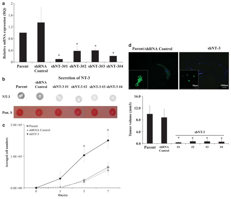
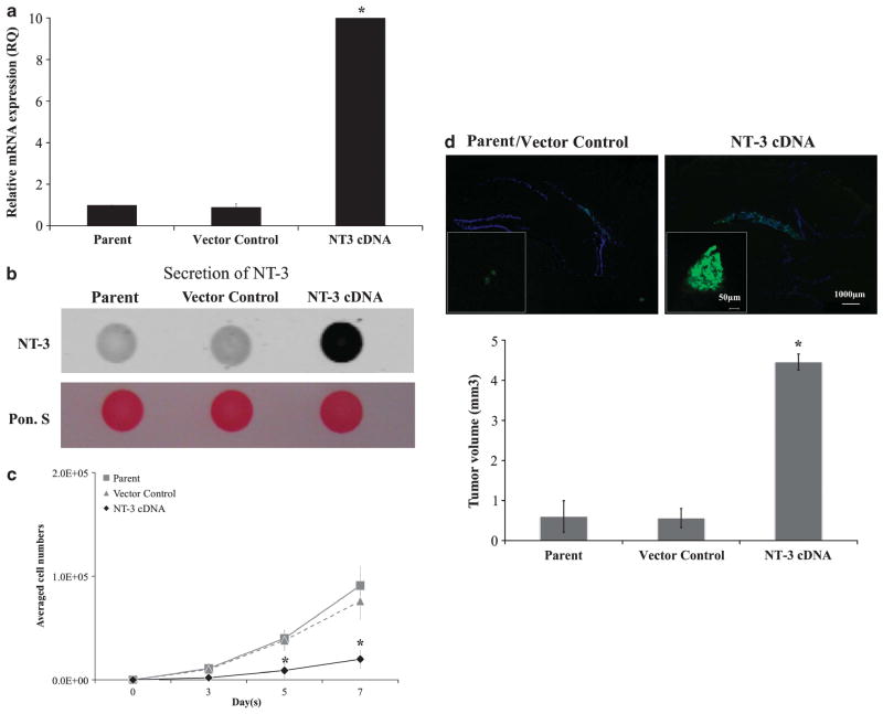
Metastasis, which remains incompletely characterized at the molecular and biochemical levels, is a highly specific process. Despite the ability of disseminated cancer cells to intravasate into distant tissues, it has been long recognized that only a limited subset of target organs develop clinically overt metastases. Therefore, subsequent adaptation of disseminated cancer cells to foreign tissue microenvironment determines the metastatic latency and tissue tropism of these cells. As a result, studying interactions between the disseminated cancer cells and the adjacent stromal cells will provide a better understanding of what constitutes a favorable or unfavorable microenvironment for disseminated cancer cells in a tissue-specific manner. Previously, we reported a protein signature of brain metastasis showing increased ability of brain metastatic breast cancer cells to counteract oxidative stress. In this study, we showed that another protein from the brain metastatic protein signature, neurotrophin-3 (NT-3), has a dual function of regulating the metastatic growth of metastatic breast cancer cells and reducing the activation of immune response in the brain. More importantly, increased NT-3 secretion in metastatic breast cancer cells results in a reversion of mesenchymal-like (EMT) state to epithelial-like (MET) state and vice versa. Ectopic expression of NT-3 in EMT-like breast cancer cells reduces their migratory ability and increases the expression of HER2 (human epidermal growth factor receptor 2) and E-cadherin at the cell-cell junction. In addition, both endogenous and ectopic expression of NT-3 reduced the number of fully activated cytotoxic microglia. In summary, NT-3 appears to promote growth of metastatic breast cancer cells in the brain by facilitating the re-epithelialization of metastatic breast cancer cells and downmodulating the cytotoxic response of microglia. Most importantly, our results provide new insights into the latency and development of central nervous system macrometastases in patients with HER2-positive breast tumors and provide mechanistic rationale to target HER2 signaling for HER2-positive breast cancer brain metastasis.


Conflict of interest statement

CONFLICT OF INTEREST

The authors declare no conflict of interest.

Figures

Figure 1
Figure 1
Expression profiles of NTs and neurotrophin TRKs in brain selective and non-brain selective breast cancer cell lines. (a) mRNA expression of NTs and TRKs in brain selective (MDA-MB 361 and BCM2 BrainG2) and non-brain selective (MDA-MB 231 and BCM2) breast cancer cells. Quantitative real-time PCR was used to compare the mRNA levels of these cell lines. MDA-MB 231 was used as the reference cell line to derive relative quantification (RQ) values for mRNA expression. (b) Secretion of NT-3 in the conditioned media by the dot blot analysis. A total of 20 μg of proteins from the conditioned media of each cell line were spotted on the nitrocellulose membrane. Antibody against NT-3 was used to detect the presence of secreted NT-3 in the conditioned media. Ponceau S staining was used to show equal loading of proteins from the conditioned media. Triplicate dots per cell line were used for quantitative analysis (see Materials and methods for quantitative analysis). BCM2 NT-3 intensity was used as the reference to derive relative fold difference of secreted NT-3 in other cell lines. (c) Growth response elicited by NT-3 in the serum-free culture media. The growth response was measured by the cell proliferation assay. Values in parenthesis are the concentrations of NT-3 used in this assay in ng/ml. *Statistically significant values (P<0.05). A full colour version of this figure is available at the Oncogene journal online.
Figure 2
Figure 2
Detection of NT-3 protein in brain metastases from breast cancer patients. (a) × 100 magnification of the brain metastasis tissue section with the isotype-matched negative control antibody staining. (b) ×100 magnification of the brain metastasis tissue section with the NT-3 antibody staining. (c) ×200 magnification of the brain metastasis tissue section with the hematoxylin and eosin (H&E) staining. (d) ×400 magnification of the brain metastasis tissue section stained with the NT-3 antibody. (e) ×400 magnification of the primary breast tumor tissue section stained with the NT-3 antibody. (f) ×200 magnification of the normal brain section stained with the NT-3 antibody.
Figure 3
Figure 3
Reduced NT-3 expression in brain selective breast cancer cells increase cell proliferation in vitro but decrease the metastatic growth of these cells in vivo. (a) mRNA expression of NT-3 in MDA-MB 361 breast cancer cells without transfection (Parent), transfected with scramble shRNA (shRNA Control), and transfected with shRNAs against NT-3 (shNT-3 nos. 1–4). (b) Dot blot analysis of NT-3 protein in the condition media. (c) Proliferation of cells with or without shNT-3. (d) Representative images and tumor volumes of metastatic brain lesions derived from cells transfected with shRNA control (left) or the shRNA against NT-3 (right; n = 6 in each group). *Statistically significant values (P<0.05).
Figure 4
Figure 4
Increased NT-3 expression in non-brain selective breast cancer cells decrease cell proliferation in vitro but increase the metastatic growth of these cells in vivo. (a) mRNA expression of NT-3 in MDA-MB 231 breast cancer cells without transfection (Parent), transfected with an empty vector (Vector Control) and transfected with NT-3 cDNA (NT-3 cDNA). (b) Dot blot analysis of NT-3 protein in the condition media. (c) Proliferation of cells with or without overexpressing NT-3 cDNA. (d) Representative images and tumor volumes of metastatic brain lesions derived from cells transfected with an empty vector (left) or the NT-3 cDNA (right) (n = 6 in each group). *Statistically significant values (P<0.05).
Figure 5
Figure 5
Organotypic cultures of metastatic breast cancer cells. (a) Colonization of metastatic breast cancer cells with endogenous NT-3 or transfected with shNT-3 or NT-3 cDNA on organotypic brain slice cultures. Green fluorescence proteins expressed in the cells were used to visualize the colony formation and quantify the growth of the colonies using the scanning confocal microscope. (b) Quantification of GFP colonies by NIH ImageJ software at different time points. *Statistically significant values (P<0.05).
Figure 6
Figure 6
Genetic, morphological and functional changes of metastatic breast cancer cells with increased or decreased NT-3 expression. (a) Relative mRNA changes of luminal and myoepithelial markers in brain selective breast cancer cells (MDA-MB 361 and MDA-MB 231 + NT-3 cDNA) and non-brain selective breast cancer cells (MDA-MB 231 and MDA-MB 361 + NT-3shRNA). (b) Immunostaining of HER2 and Keratin8/ 18 in the metastatic lesions of brains implanted with either brain selective or non-brain selective breast cancer cells. (c) Immunostaining of E-cadherin and Snail in the metastatic lesions of brains implanted with either brain selective or non-brain selective breast cancer cells. (d) Wound-healing assay to evaluate the migratory ability of brain selective and non-brain selective breast cancer cells. (e) A representative gross image of spontaneous brain metastasis with disruption of blood–brain barrier (left panel) and a confocal image of brain metastasis GFP colonies in a brain section (right panel). (f) Immunostaining of NT-3 and E-cadherin in lesions of spontaneous brain metastasis. The stem-like subpopulation was engineered to express GFP for visualization.
Figure 7
Figure 7
HER2 inhibitors are effective in suppressing HER2 phosphorylation of brain selective and non-brain selective breast cancer cells in vitro and reducing the metastatic of these cells in vivo. (a) Changes of TrkC (NT-3 high-affinity receptor) and HER2 mRNA and protein levels in brain selective and non-brain selective breast cancer cells. (b) Western blot analysis of total HER2 and phosphorylated HER2 (Y877) in the presence of exogenous NT-3 with or without the HER2 small-molecule inhibitors (Lapatinib and AG874). (c) Dose-response curve of brain selective and non-brain selective breast cancer cells against HER2 small-molecule inhibitors. (d) Changes in metastatic growth of brain selective breast cancer cells in vivo after the Lapatinib and AG874 treatments. *Statistically significant values (P<0.05).
Figure 8
Figure 8
Microglia activation in the metastatic lesions of breast cancer cells in vivo. (a) Staining of microglia surrounding and infiltrating the metastatic lesions derived from brain selective and non-brain selective breast cancer cells. Iba-1 staining was used to visualize activated microglia. (b) Quantifications of the percentage of activated microglia and tumor volumes in the metastatic lesions derived from brain selective and non-brain selective breast cancer cells.

Similar articles

Cited by

References

    1. Welch DR, Steeg PS, Rinker-Schaeffer CW. Molecular biology of breast cancer metastasis. Genetic regulation of human breast carcinoma metastasis. Breast Cancer Res. 2000;2:408–416. - PMC - PubMed
    1. Weil RJ, Palmieri DC, Bronder JL, Stark AM, Steeg PS. Breast cancer metastasis to the central nervous system. Am J Pathol. 2005;167:913–920. - PMC - PubMed
    1. Price JE. Metastasis from human breast cancer cell lines. Breast Cancer Res Treat. 1996;39:93–102. - PubMed
    1. Minn AJ, Gupta GP, Siegel PM, Bos PD, Shu W, Giri DD, et al. Genes that mediate breast cancer metastasis to lung. Nature. 2005;436:518–524. - PMC - PubMed
    1. Lu X, Kang Y. Organotropism of breast cancer metastasis. J Mammary Gland Biol Neoplasia. 2007;12:153–162. - PubMed

Publication types