. 2012 Nov;117(5):1080-90.
doi: 10.1097/ALN.0b013e31826f8d86.

Propofol at clinically relevant concentrations increases neuronal differentiation but is not toxic to hippocampal neural precursor cells in vitro

Affiliations

Propofol at clinically relevant concentrations increases neuronal differentiation but is not toxic to hippocampal neural precursor cells in vitro

Jeffrey W Sall et al. Anesthesiology. 2012 Nov.

Abstract

Background: Propofol in the early postnatal period has been shown to cause brain cell death. One proposed mechanism for cognitive dysfunction after anesthesia is alteration of neural stem cell function and neurogenesis. We examined the effect of propofol on neural precursor or stem cells (NPCs) grown in vitro.

Methods: Hippocampal-derived NPCs from postnatal day 2 rats were exposed to propofol or Diprivan. NPCs were then analyzed for bromodeoxyuridine incorporation to measure proliferation. Cell death was measured by lactate dehydrogenase release. Immunocytochemistry was used to evaluate the expression of neuronal and glial markers in differentiating NPCs exposed to propofol.

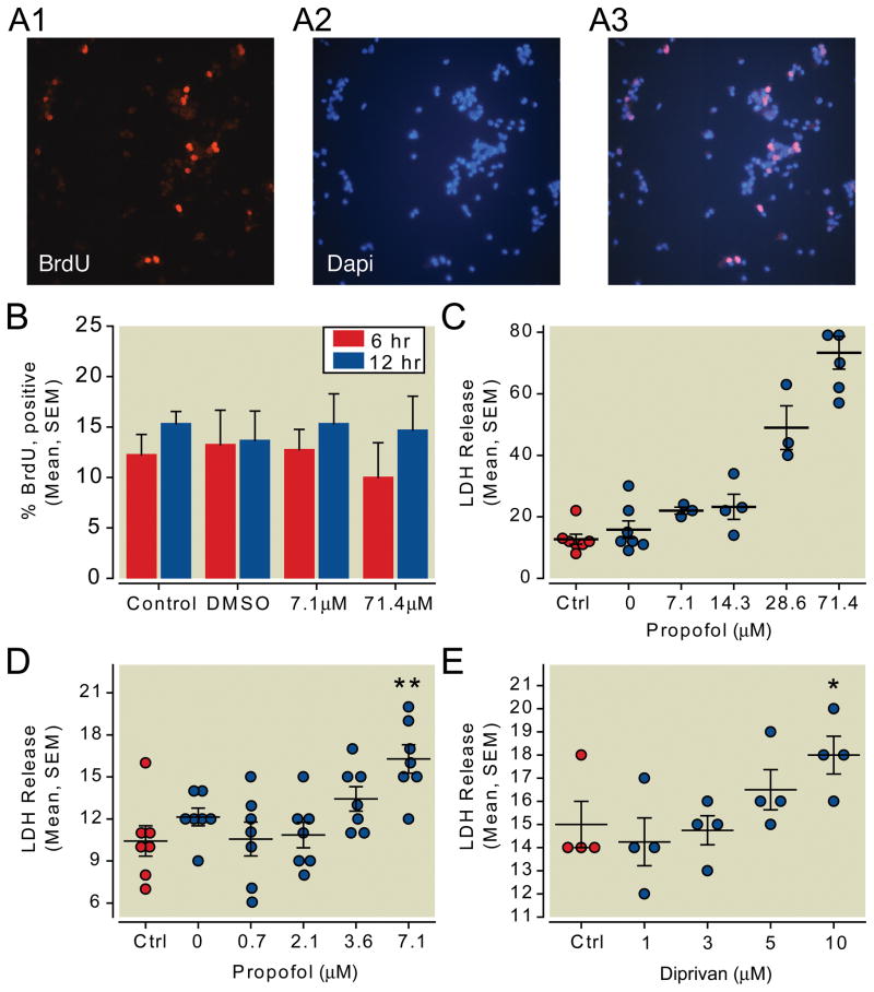
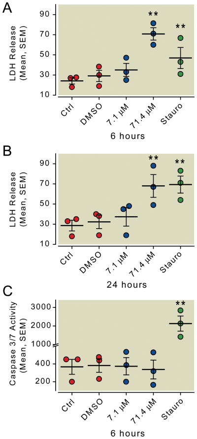
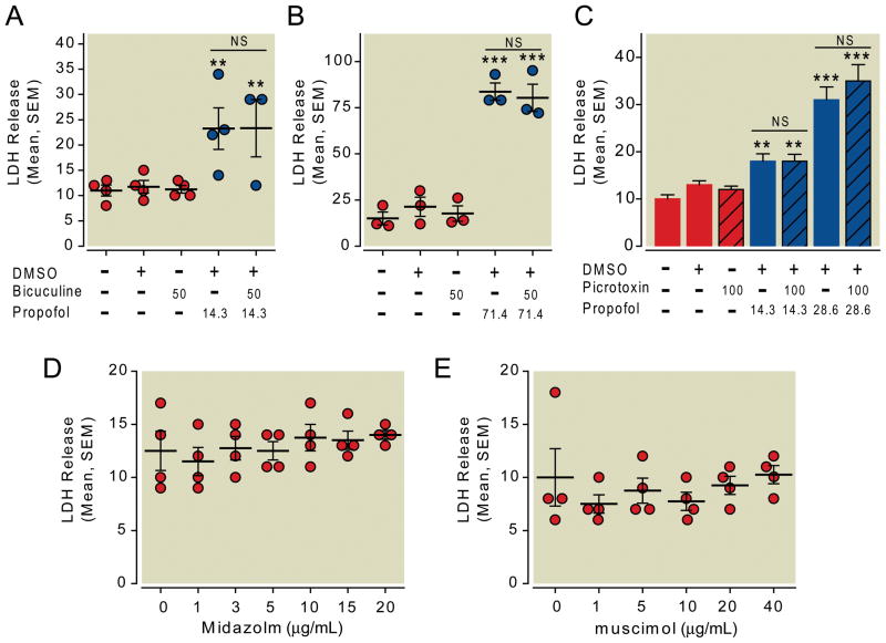
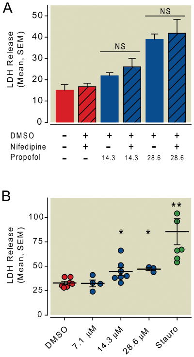
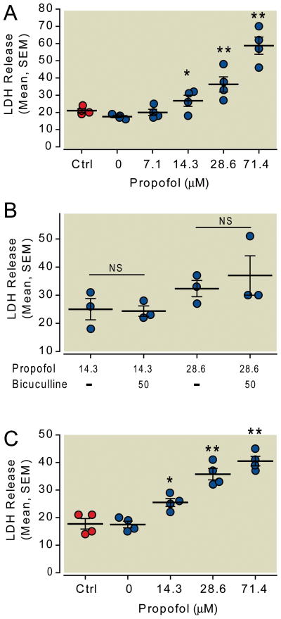
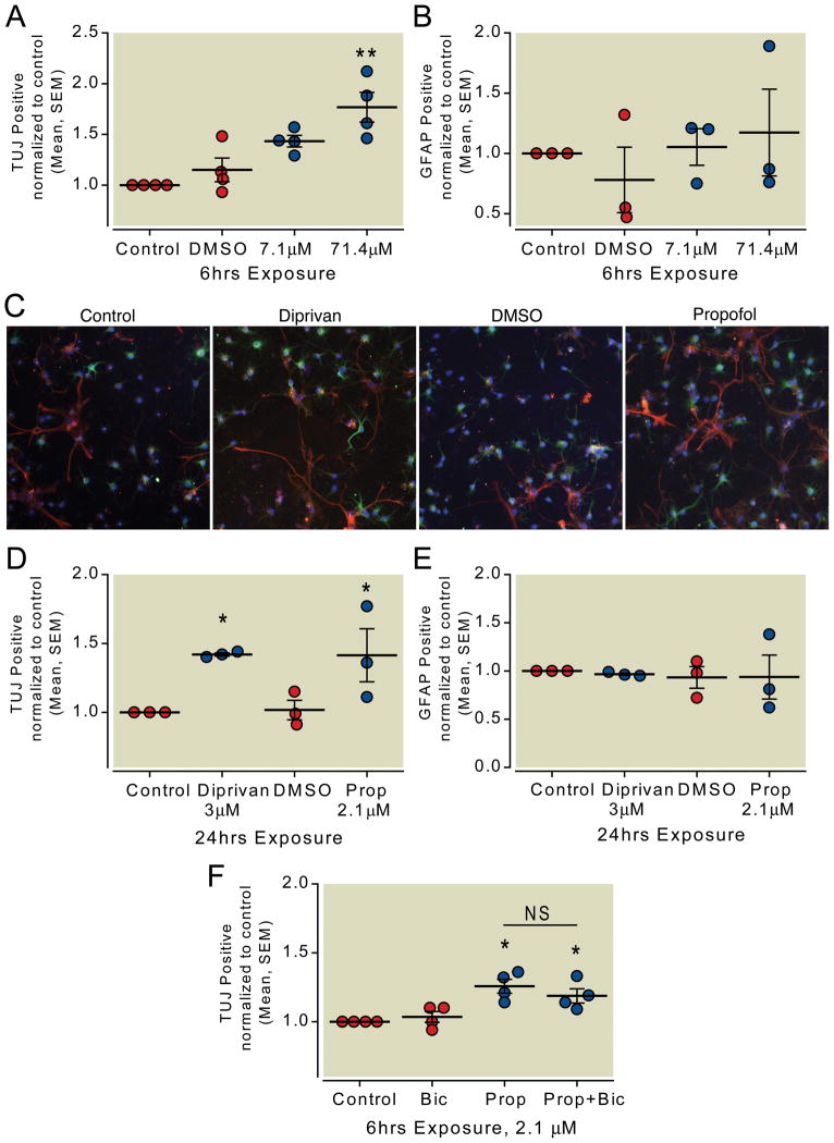
Results: Propofol dose dependently increases the release of lactate dehydrogenase from NPCs under both proliferating and differentiating conditions at supraclinical concentrations (more than 7.1 µM). Both Diprivan and propofol had the same effect on NPCs. Propofol-mediated release of lactate dehydrogenase is not inhibited by blocking the γ-aminobutyric acid type A receptor or extracellular calcium influx and is not mediated by caspase-3/7. Direct γ-aminobutyric acid type A receptor activation did not have the same effect. In differentiating NPCs, 6 h of propofol at 2.1 µM increased the number neurons but not glial cells 4 days later. Increased neuronal differentiation was not blocked by bicuculline.

Conclusions: Only supraclinical concentrations of propofol or Diprivan kill NPCs in culture by a non-γ-aminobutyric acid type A, noncaspase-3 mechanism. Clinically relevant doses of propofol increase neuronal fate choice by a non-γ-aminobutyric acid type A mechanism.


Figures

Fig. 1
Fig. 1
Propofol did not change the proportion of cells in S-phase, but did cause dose dependent toxicity in proliferating neural precursor or stem cells (NPCs). (A)NPCs were given a pulse of BrdU after six or 24 hours exposure to propofol then fixed for immunocytochemistry with anti 5-bromo-2′-deoxyuridine antibody(A1) or 4′,6-diamidino-2-phenylindole(A2). Overlayed images (A3) were then photographed and the nuclei were counted. (B)The number of cells that incorporated BrdU was not different in cells treated with propofol vs dimethyl sulfoxide carrier only. (C)NPCs were exposed to increasing concentrations of propofol for 6 hours then assayed for lactate dehydrogenase release into the media. All values are relative to complete lysis of cells which was set at 100%. Increasing the concentration of propofol increased the amount of lactate dehydrogenase released in the media. (D)The low end of the dose response curve was further investigated and 7.1μM propofol was found to be different from 0μM propofol (carrier only)(Dunnett’s post test). (E)Diprivan, a clinically used formulation of propofol, produced a similar result when compared to control (Dunnett’s post test). Ctrl=control, LDH=Lactate Dehydrogenase, DMSO=dimethyl sulfoxide,* P < 0.05, ** P < 0.01.
Fig. 2
Fig. 2
Toxicity is not mediated by caspase 3/7 activation. Neural precursor cells grown in proliferation medium were exposed to propofol or staurosporine as a positive control for caspase activation. Like propofol, staurosporine caused some lactate dehydrogenase release after 6 hours (A), and more lactate dehydrogenase release 24 hours later (B). However, only staurosporine induced caspase 3/7 activation in neural precursor cells (C)(Dunnett’s post test, ** P < 0.01). LDH=lactate dehydrogenase, Ctrl=control, DMSO=dimethyl sulfoxide
Fig. 3
Fig. 3
Neural precursor cells grown in culture express the γ-aminobutyric acid type A (GABAA) receptor. Western blot analysis reveals that 55 kD GABAA receptor alpha-1 subunit is expressed in whole brain but not in independently isolated cultured neural stem cell lines (A). The GABAA receptor beta-3 subunit was found in all stem cell lines that were analyzed as well as in whole brain (B). Glyceraldehyde phosphate dehydrogenase (GAPDH) was used as a control.
Fig. 4
Fig. 4
Propofol toxicity is not mediated by γ-aminobutyric acid type A (GABAA) receptor activation. Neural precursor cells in proliferation media were exposed to propofol in the presence or absence of the GABAA receptor antagonists bicuculline or picrotoxin. Six hours of 14.3μM (A) or 71.4μM (B) propofol induced lactate dehydrogenase (LDH) release that was not inhibited by addition of bicuculline. Similarly, the channel blocker picrotoxin did not block LDH release after propofol exposure for 6 hours(C). Treating neural precursor cells with GABAA receptor agonists midazolam (D) or muscimol (E) did not cause release of LDH from neural precursor cells over a wide range of concentrations (Bonferroni’s post test vs control or treatment pair). * P < 0.05, ** P < 0.01, *** P < 0.001, NS = not significant, DMSO = dimethyl sulfoxide.
Fig. 5
Fig. 5
Propofol toxicity is not mediated by extracellular calcium. Neural precursor cells growing in proliferation media were exposed to propofol in the presence or absence of the calcium channel blocker nifedipine. (A) Six hours of propofol exposure induced lactate dehydrogenase (LDH) release. Propofol mediated LDH release was not inhibited by addition of 10μM nifedipine (Bonferroni’s post test). (B) Neural precursor cells grown in proliferation media were transferred to calcium free media and treated with various concentrations of propofol. The media change caused an elevated baseline value but propofol exposure still led to increased LDH release into the media (Bonferroni’s post test vs dimethyl sulfoxide or DMSO control). The positive control staurosporine which induced caspase mediated cell death also caused cell death and LDH release under these calcium free conditions. NS = not significant, * P < 0.05.
Fig. 6
Fig. 6
Neural precursor cells in differentiation medium are not protected from propofol toxicity. (A) Neural precursor cells growing in proliferation medium were switched to differentiation conditions at the time of propofol exposure. Increasing doses of propofol exposure for six hours under differentiating conditions caused cell damage and release of lactate dehydrogenase (LDH) into the medium (Dunnett’s multiple comparison vs 0μM). (B) LDH release after six hours of propofol was not inhibited by addition of bicuculline to the medium (Bonferroni’s multiple comparison). (C) Neural precursor cells were switched to differentiation conditions 24 hours prior to addition of propofol. Increasing doses of propofol still caused a rise in LDH release (Dunnett’s multiple comparison vs 0μM). Ctrl = control, NS = not significant, * P < 0.05, ** P < 0.01.
Fig. 7
Fig. 7
Propofol increases expression of neuron specific Class III β-tubulin (Tuj1) in differentiating precursor cells. Neural precursor cells were plated in differentiation medium with propofol for six hours then washed to remove propofol and serum. Four days later the expression of Tuj1 and glial fibrillary acidic protein (GFAP) was evaluated by immunocytochemistry using fluorescence microscopy. (A) Neuronal precursors differentiated in the presence of propofol were more likely to express the early neuronal marker Tuj1 (Dunnett’s multiple comparison vs Control). (B) No difference was seen in expression of GFAP. (C) Representative photomicrographs of neural precursor cells that were differentiated under serum free conditions with 3μM Diprivan or 2.1μM propofol for 24 hours then washed to remove the propofol. After four days cells were evaluated for expression of Tuj1 (green) or GFAP (Red) and 4′,6-diamidino-2-phenylindole (Blue) to mark the nuclei. (D) Both the clinical preparation, Diprivan, and propofol in dimethyl sulfoxide increased the number of cells expressing Tuj1 four days later (Bonferroni’s post test). (E) no difference was seen in the number of cells expressing GFAP. (F) Neuronal precursor cells were exposed to 2.1μM propofol for six hours in the presence or absence of bicuculline. Media was changed to remove propofol or bicuculline and cells were allowed to differentiate. Four days after low dose propofol exposure Tuj1 expression was increased and this was not blocked by addition of bicuculline to the medium (Bonferroni’s multiple comparison). Ctrl = control, DMSO = dimethylsulfoxide, Prop = propofol, Bic = bicuculline, NS = not significant, * P < 0.05, ** P < 0.01.

Similar articles

Cited by

References

    1. Cattano D, Young C, Straiko MM, Olney JW. Subanesthetic doses of propofol induce neuroapoptosis in the infant mouse brain. Anesth Analg. 2008;106:1712–4. - PubMed
    1. Milanovic D, Popic J, Pesic V, Loncarevic-Vasiljkovic N, Kanazir S, Jevtovic-Todorovic V, Ruzdijic S. Regional and temporal profiles of calpain and caspase-3 activities in postnatal rat brain following repeated propofol administration. Dev Neurosci. 2010;32:288–301. - PMC - PubMed
    1. Pesic V, Milanovic D, Tanic N, Popic J, Kanazir S, Jevtovic-Todorovic V, Ruzdijic S. Potential mechanism of cell death in the developing rat brain induced by propofol anesthesia. Int J Dev Neurosci. 2009;27:279–87. - PMC - PubMed
    1. Bercker S, Bert B, Bittigau P, Felderhoff-Muser U, Buhrer C, Ikonomidou C, Weise M, Kaisers UX, Kerner T. Neurodegeneration in newborn rats following propofol and sevoflurane anesthesia. Neurotox Res. 2009;16:140–7. - PubMed
    1. Trapani G, Altomare C, Liso G, Sanna E, Biggio G. Propofol in anesthesia. Mechanism of action, structure-activity relationships, and drug delivery. Curr Med Chem. 2000;7:249–71. - PubMed

Publication types

MeSH terms