. 2012 Nov;13(11):1083-91.
doi: 10.1038/ni.2428. Epub 2012 Sep 23.

The proto-oncogene MYC is required for selection in the germinal center and cyclic reentry

Affiliations

The proto-oncogene MYC is required for selection in the germinal center and cyclic reentry

David Dominguez-Sola et al. Nat Immunol. 2012 Nov.

Abstract

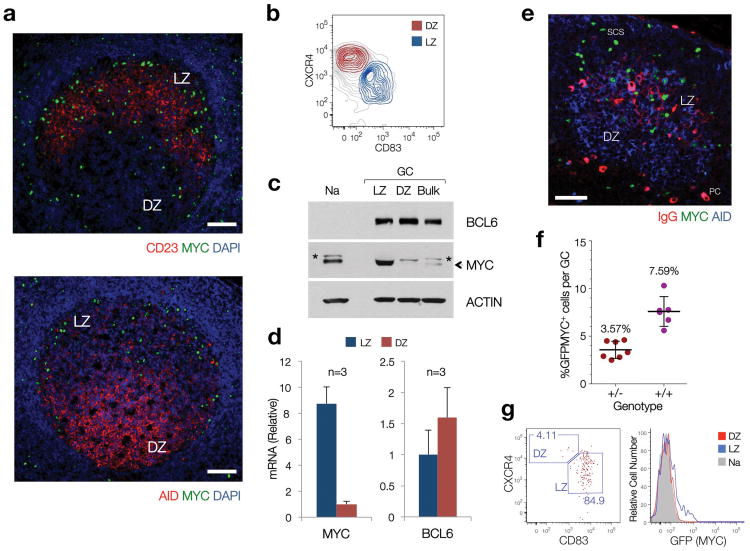
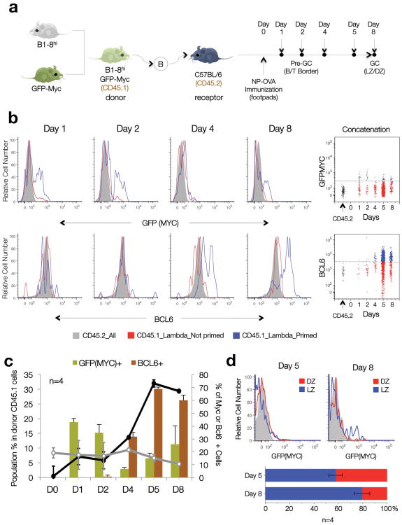
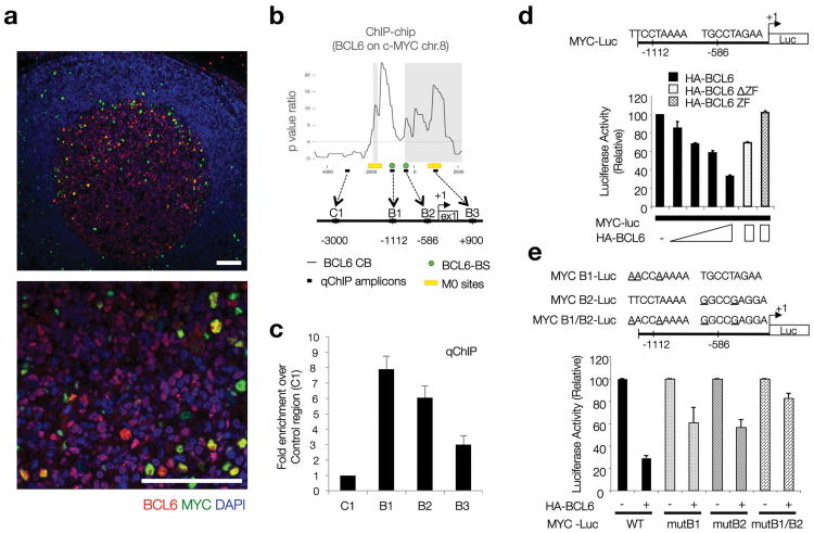
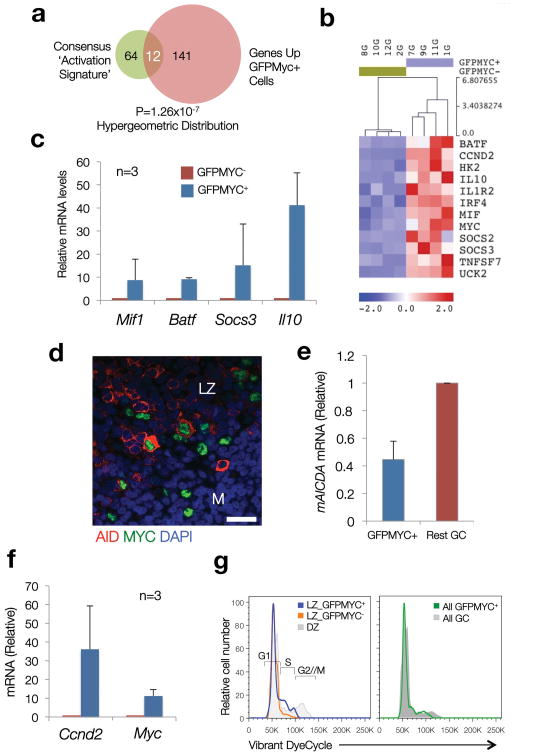
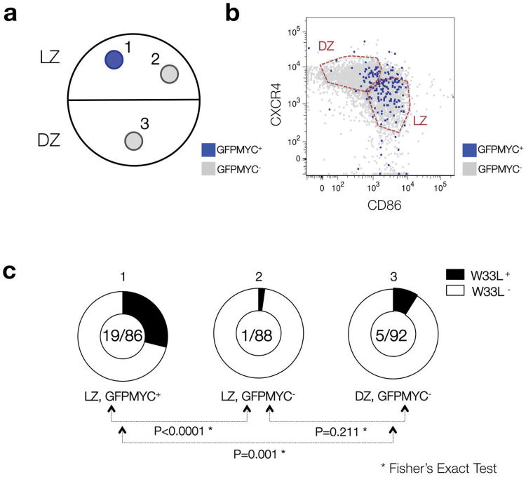
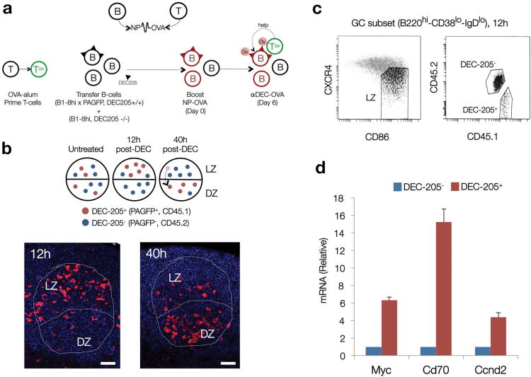
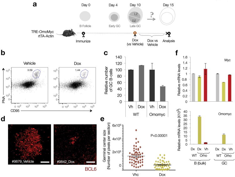
After antigenic challenge, B cells enter the dark zone (DZ) of germinal centers (GCs) to proliferate and hypermutate their immunoglobulin genes. Mutants with greater affinity for the antigen are positively selected in the light zone (LZ) to either differentiate into plasma and memory cells or reenter the DZ. The molecular circuits that govern positive selection in the GC are not known. We show here that the GC reaction required biphasic regulation of expression of the cell-cycle regulator c-Myc that involved its transient induction during early GC commitment, its repression by Bcl-6 in DZ B cells and its reinduction in B cells selected for reentry into the DZ. Inhibition of c-Myc in vivo led to GC collapse, which indicated an essential role for c-Myc in GCs. Our results have implications for the mechanism of GC selection and the role of c-Myc in lymphomagenesis.


Figures

Figure 1
Figure 1. A subset of LZ GC B cells express MYC under physiologic conditions
(a) Immunofluorescence staining, paraffin-embedded sections of reactive human lymph nodes. CD23, expressed in Follicular Dendritic cells (FDC), highlights the boundaries of the LZ. AID is used as a DZ marker, as previously reported, . Scale Bars = 200μm. (b) Sorting profiles of LZ and DZ GC B cell subpopulations in human tonsils (See Supplementary Fig. 1) (c) Immunoblot of populations shown in (b). (Na), Naive B cells. (Bulk), bulk CD77+ GC B cells, isolated as in. The asterisk denotes a non-specific band. Actin is used as loading control. (d) Quantitative RT-PCR for MYC and BCL6 mRNA levels in LZ and DZ B cell pools. Average of 3 independent cell pools per population (n=3). Error bars, standard deviation (SD). (e) Immunofluorescence staining, paraffin-embedded section, mouse lymph node (12 days after SRBC immunization). AID highlights the GC (DZ). IgG highlights the FDC network (i.e. LZ). Scale bar = 50μm. PC, plasma cell. SCS, subcapsular sinus (f) Number of MYC+ B cells, as assessed by flow cytometry (GFP=MYC) in LZ and DZ GC B cell subsets from GFPMYC mice (12 days post-immunization). (g) Distribution of GFPMYC+ GC B cells among LZ and DZ subsets. Left panel, dot plot analysis. Right panel, histogram shows the relative GFPMYC fluorescence intensities within these cell subsets. Shadowed histogram, Naive B cells (Na), used as a reference.
Figure 2
Figure 2. Alternating peaks of MYC and BCL6 expression during T cell-dependent antigen responses and GC formation
(a) Diagrammatic representation of the experimental approach. (b) Representative histogram plots depict the distribution of GFPMYC (top) and BCL6 (bottom) protein expression in primed, Igλ, CD45.1+ (blue profiles); non-primed, Igλ, CD45.1+ B1-8hi B cells (red profiles), and CD45.2 host B cells (shadowed histograms) at different time points after NP-OVA immunization (see also Supplementary Fig. 2). The bidimensional dotplots shown on the right panels summarize the data by concatenating all samples (n=4). Dashed lines mark the background levels for GFPMYC and BCL6 fluorescent signals. (c) Bar and line graphs summarize the temporal evolution in cell numbers (black and grey lines, left axis) and GFPMYC and BCL6 protein expression (right axis), in the antigen-primed B1-8hi B cell population during GC formation. Error bars=SD (n=4). (d) Topographic distribution of GFPMYC positive, primed B cells, within each GC compartment (LZ, DZ) at Day 5 and 8 post-immunization (flow cytometry). The average number of GFPMYC positive cells in each compartment is summarized in the graph below (n=4). Error bars=SD.
Figure 3
Figure 3. BCL6 represses MYC protein expression in DZ GC B cells
(a) Immunofluorescence co-staining for MYC and BCL6 in human reactive lymph nodes. BCL6 (red) highlights the boundaries of the GC. DAPI (blue), nuclear counterstain. Scale bars= 200μm. (b) BCL6 chromatin binding profile (ChIP-chip) at the MYC locus in human CD77+ GC B cells. The diagram shows the organization of this locus in the region around Exon1 and the transcription start site (+1). B6BS and M0 potential BCL6 binding sites are based on previously defined consensus. The qChIP amplicons referred to in panel (c) are shown here as black boxes (B1-3, C1). Raw data corresponding to the ChIP-chip analysis at the MYC locus is provided in Supplementary Table 1, available online. (c) Quantitative PCR on DNA isolated from an independent BCL6 chromatin immunoprecipitation assay in CD77+ GC B cells. Fold enrichment refers to the ‘normalized’ binding at each region, relative to C1 (arbitrarily set to 1). ‘Normalized binding’ measures the relative enrichment in BCL6 immunoprecipitates over background (i.e. species or isotype matched irrelevant antibody). Average of 3 technical replicates (Error bar=SD). (d) Dual-Luciferase reporter assay, HEK293T cells. Effects of WT BCL6 or two defective truncations (as indicated) on the 1.2 Kb upstream MYC regulatory region depicted in the diagram, which includes two BCL6 potential binding sites. Average of 2 technical replicates from a representative experiment (Error bar=SD). (e) Same assay as in panel (d), but using Luciferase reporter constructs where point mutations on each putative BCL6 consensus site were introduced, as indicated. Average of 2 technical replicates from a representative experiment (Error bar=SD).
Figure 4
Figure 4. Coordinated upregulation of immune activation signatures and cell cycle entry genes in GFPMYC+ GC B cells
(a) A consensus ‘Activation Signature’, integrated by genes common to two or more published ‘immune activation’ signatures, was built (76 genes, details in Supplementary Table 4). The overlap between this signature and that of GFPMYC+ GC B cells (Venn diagram) was determined using a hypergeometric distribution (=12 genes). (Normalized Enrichment Score (NES)=2.34 (P val<0.00001; FDR=0%) using the GSEA algorithm). (b) Expression profile of the 12 consensus ‘immune activation’ genes enriched in GFPMYC+ cells. (c) Relative mRNA levels of selected ‘immune activation’ genes in GFPMYC+ cells, average of 3 independent cell pools (2 mice per pool). Error bars=SD. (d) Co-expression of AID (red) and MYC (green) in murine GC B cells (immunofluorescence, murine lymph node; DAPI (blue), nuclear counterstain). Scale bar, 20 μm. (e) Relative Aicda (AID) mRNA levels in GC B cell subsets isolated from GFPMYC mice, 12 days after SRBC immunization. Average of 2 independent experiments. Error bars, SD. (f) Relative Ccnd2 mRNA levels in GFPMYC+ and GFPMYC- populations (qRT-PCR). Average of 3 independent cell pools (2 mice per pool). (g) Cell cycle profile analysis in LZ and DZ GC B cells. Surface CXCR4-CD86 define the LZ and DZ subpopulations. These histograms correspond to one mouse, representative of 3. Cell cycle phases are defined based on DNA content, adjusted to a Watson-Pragmatic model. Right panel, cell cycle distribution in bulk GFPMYC+ GC B cells, as compared to whole GC B cells.
Figure 5
Figure 5. The B cell receptor (BCR) repertoire of GFPMYC positive GC B cells is enriched in high affinity variants
(a) Diagrammatic representation of the 3 different populations purified from GC B cell pools of GFPMYC mice, 9 days after immunization with NP-KLH. (b) Distribution of GFPMYC+ and GFPMYC- subpopulations within these compartments. The detailed gating strategy to isolate the GC cell subsets is described in Supplementary Figure 1. (c) Doughnut charts show the fraction of sequenced Vh186.2 segments (CDR1 region) with a W33L mutation among all sequenced segments (W33L mutants/Total segments). See Supplementary Table 5 for additional details. The results correspond to the pooled data of two independent experiments, 2-3 mice analyzed per experiment. P value calculations are based on a Fisher's exact test.
Figure 6
Figure 6. Access to T cell help triggers MYC expression prior to DZ re-entry
(a) Diagrammatic representation of the experimental strategy. NP haptens are detected by B cells. Ovalbumin peptide conjugates (OVA), by T cells. DEC-205+ (CD205+, Ly75) cells are represented with triangular segments on their surface. Red cells, NP primed. OV labeled circles, OVA. (b) Top panel, expected dynamics and topographical distribution of DEC-205+ (PAGFP+) cells before (untreated) and after injection of OVA conjugated anti-DEC-205 antibodies (αDEC-OVA). Red cells, DEC-205+. Blue cells, DEC-205-. Bottom panels, immunofluorescence analysis on representative paraffin-embedded sections of popliteal lymph nodes after αDEC-OVA injection, at the indicated time points, to detect PAGFP+ (DEC-205+) B cells within B220+ GC compartments (outlined). LZ (Light Zone), DZ (Dark Zone). Scale bars = 50 μm. (c) Gating strategy used to isolate DEC-205+ and DEC-205- GC B cells from the LZ of the experimental mouse cohort 12 hours after αDEC-OVA injection. (d) Quantitative analysis for Myc, Cd70 and Ccnd2 mRNA levels in DEC-205+ and DEC-205- populations, isolated as shown in panel (c). Shown is one representative experiment (n=2). Average of 3 technical replicates +/- SD). See also Supplementary Fig. 5.
Figure 7
Figure 7. MYC biological activity is required for normal GC maintenance
(a) Diagrammatic representation of the experimental approach, as described in the main text, and the expected dynamics in T-dependent antigen responses and GC formation (Dox=Doxycycline) (SRBC= Sheep Red Blood Cells) (b) Representative Dot Plots on B cell splenic pools from Control and Dox-induced TRE-Omomyc rtTA-actin mice. The CD95hiPNAhi gate defines the GC population (blue outline). The number indicates the percentage of GC B cells. 2 mice per condition were analyzed. (c) Bar graphs show the average percentage of GC B cells in each group of samples (n=2 per condition). Error bars=SD. (d) Representative images of GC in paraffin-embedded sections of spleens from control (Vehicle) and Dox-induced (Dox) mice. BCL6 stain highlights GC B cells. Scale bars= 50 μm.(e) GC size distribution (surface) in control (Vhc) and Dox-induced (Dox) Omomyc mice (2 mice per condition pooled). Each marker corresponds to a single GC, its surface represented in number of pixels (∼50-55 GCs per mouse, see Methods online). Horizontal lines highlight average values. Error bars=SE. P value calculated using a two-tailed Student's T-test, unequal variance. (f) Quantitative analysis of Myc and Omomyc mRNA levels in bulk B cell pools and GC fractions. mRNA obtained from SRBC immunized wild-type (WT) mice was used as a reference control. Data analysis was performed by the ΔΔCt method, upon normalization to the geometric mean of Ct values for 3 different housekeeping genes (Gapdh, Actb and Hprt1). Bar graphs correspond to the average of 2 mice (Error bars=SD).

Comment in

  • The case of the missing c-Myc.
    Shaffer AL 3rd, Staudt LM. Shaffer AL 3rd, et al. Nat Immunol. 2012 Nov;13(11):1029-31. doi: 10.1038/ni.2455. Nat Immunol. 2012. PMID: 23080195 No abstract available.

References

    1. Allen CD, Okada T, Cyster JG. Germinal-center organization and cellular dynamics. Immunity. 2007;27:190–202. - PMC - PubMed
    1. MacLennan IC. Germinal centers. Annu Rev Immunol. 1994;12:117–139. - PubMed
    1. Victora GD, Nussenzweig MC. Germinal centers. Annu Rev Immunol. 2012;30:429–457. - PubMed
    1. Victora GD, et al. Identification of human germinal center light and dark zone cells and their relationship to human B cell lymphomas. Blood. 2012 - PMC - PubMed
    1. Allen CD, Okada T, Tang HL, Cyster JG. Imaging of germinal center selection events during affinity maturation. Science. 2007;315:528–531. - PubMed

Publication types

MeSH terms

Substances

Associated data