

Clinical and radiological profile of ameloblastic fibro-odontoma: an update on an uncommon odontogenic tumor based on a critical analysis of 114 cases
- PMID: 23001451
- PMCID: PMC3597150
- DOI: 10.1007/s12105-012-0397-9
Clinical and radiological profile of ameloblastic fibro-odontoma: an update on an uncommon odontogenic tumor based on a critical analysis of 114 cases
Abstract
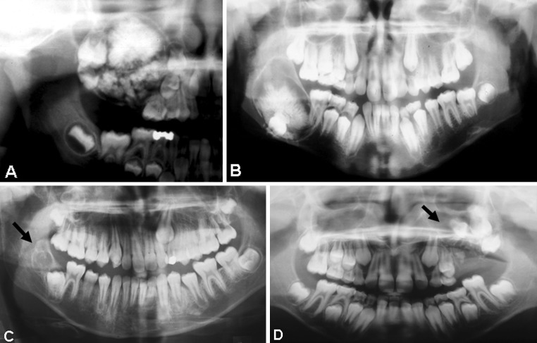
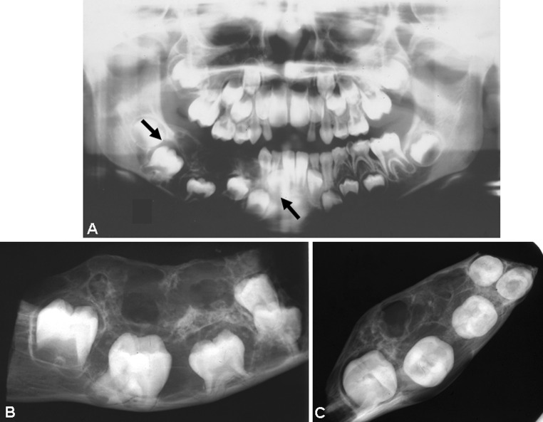
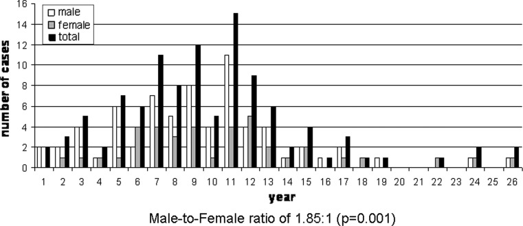
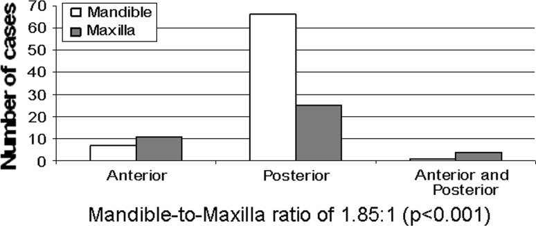
Ameloblastic fibro-odontoma is an uncommon benign tumor of the jaws that belongs to the group of mixed odontogenic tumors. The descriptions of its clinical and radiological features in the literature are not always accurate and sometimes even contradictory. The aim of the present study was to critically evaluate their clinical and radiological features as reported in the English-language literature. A total of 114 well-documented cases of ameloblastic fibro-odontomas (103 from publications and 11 of our own new cases) were analyzed. The patients' age ranged from 8 months to 26 years (mean 9.6). There were 74 (65 %) males, with a male-to-female ratio of 1.85:1 (P = 0.001). The mandible was involved in 74 (65 %) cases, and the mandible-to-maxilla ratio was 1.85:1 (P < 0.001). Nearly 80 % of the lesions were located in the posterior region of the jaws, and most (58 %) were in the posterior mandible. Radiographically, most of the lesions were unilocular and only a few (~10 %) were multilocular. Most lesions were mixed radiolucent-radiopaque, and only a few (~5 %) were radiolucent. Almost all lesions (~92 %) were associated with the crown of an unerupted tooth/teeth. This comprehensive analysis of a large number of patients with an uncommon lesion revealed that ameloblastic fibro-odontomas are significantly more common in males and in the mandible, and that multilocular lesions are uncommon. It also revealed that, based on their clinical and radiological features, some of them are probably true neoplasms while others appear to be developing odontomas (hamartomas).
Figures
References
-
- Takeda Y, Tomich CE. Ameloblastic fibro-odontoma. In: Barnes L, Eveson JW, Reichart P, Sidransky D, editors. Pathology and genetics of head and neck tumours. Lyon: IARC Press; 2005.
-
- Kramer IRH, Pindborg JJ, Shear M. Histological Typing of Odontogenic Tumours. 2. Berlin: Springer-Verlag; 1992.
-
- Pindborg JJ, Kramer IRH. Histological Typing of Odontogenic Tumours, Jaws Cysts, and Allied Lesions. Geneva: World Health Organization; 1971.
Publication types
MeSH terms
LinkOut - more resources
Full Text Sources
