

doi: 10.1002/anie.201204268.
Epub 2012 Sep 24.
Label-free microscale thermophoresis discriminates sites and affinity of protein-ligand binding
Affiliations
- PMID: 23001866
- PMCID: PMC3588113
- DOI: 10.1002/anie.201204268
Item in Clipboard
Label-free microscale thermophoresis discriminates sites and affinity of protein-ligand binding
Angew Chem Int Ed Engl.
.
Free PMC article
Abstract
Look, no label! Microscale thermophoresis makes use of the intrinsic fluorescence of proteins to quantify the binding affinities of ligands and discriminate between binding sites. This method is suitable for studying binding interactions of very small amounts of protein in solution. The binding of ligands to iGluR membrane receptors, small-molecule inhibitorss to kinase p38, aptamers to thrombin, and Ca(2+) ions to synaptotagmin was quantified.
Copyright © 2012 WILEY-VCH Verlag GmbH & Co. KGaA, Weinheim.
Figures
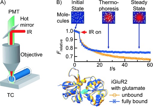
Label-free microscale thermophoresis. A) A capillary containing a protein sample with intrinsic tryptophan fluorescence is placed on a thermoelectric cooler (TC) providing a constant basis temperature. Fluorescence is excited with an UV LED and recorded with a photomultiplier tube (PMT). The solution inside the capillary is locally heated with an IR laser, which is coupled into the fluorescence microscope using an IR-reflecting “hot” mirror. B) The fluorescence of the heated spot is recorded, normalized, and plotted against time. After the IR laser is switched on at t=5 s, the fluorescence decreases as the temperature increases, and the fluorescent protein molecules move away from the heated spot because of thermophoresis. The unbound iGluR2 ligand-binding domain (yellow; PDB code 1FTO) shows stronger thermophoretic depletion than the complex with glutamate (blue; PDB code 1FTJ). This reflects the conformational change of the protein upon binding.
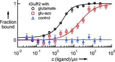
Ligand binding to membrane receptors. Binding curves are derived from the specific change in the thermophoretic mobility upon ligand titration to a constant iGluR2-LBD concentration of 2 μm . The curves show binding affinities of (835±43) nm for glutamate and (19±5) μm for glu-azo. The two agonists compete for the same binding site, as preincubation of iGluR2 with a saturating amount of glutamate prevents glu-azo binding (control).
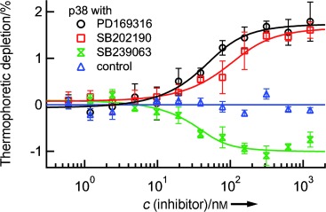
Screening of small-molecule kinase inhibitors. Three selective inhibitors were successfully tested for binding to the nonactivated form of MAP kinase p38α (c=100 nm ). Corresponding to structural differences, the binding of SB202190 and PD169316 has the opposite effect on the thermophoretic movement compared to SB239063. SB202190 binds with a KD value of (48±21) nm . The upper limits of affinity for PD169316 and SB239063 were determined as 33 nm and 20 nm , respectively. These results are in good agreement with previously reported values. Thermally denatured p38α did not show binding (control).
References
Publication types
MeSH terms
Substances
LinkOut - more resources
Full Text Sources
Other Literature Sources
Miscellaneous
